

Dealing with technical challenges in embolization of a rare aberrant left inferior bronchial artery arising from the left gastric artery in a patient with massive hemoptysis
- PMID: 30662214
- PMCID: PMC6319114
- DOI: 10.4103/ijri.IJRI_162_18
Dealing with technical challenges in embolization of a rare aberrant left inferior bronchial artery arising from the left gastric artery in a patient with massive hemoptysis
Abstract
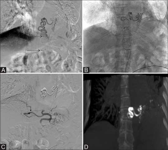
Bronchial artery embolization is an established intervention for management of recurrent massive hemoptysis in a majority of patients. The source of bleeding in a majority of cases is systemic arteries - orthotopic bronchial arteries, anomalous bronchial arteries, or nonbronchial systemic collaterals. We report a case of an aberrant left inferior bronchial artery arising from the left gastric artery (LGA) in a patient with massive hemoptysis. Such origin from infradiaphragmatic vessels and specially left gastric arteries is very rare and needs to be considered by interventional radiologists and pulmonologists in case with hemoptysis disproportionate to supply by orthotopic arteries. Technical challenges were present in the present case in the form of an aneurysm in the aberrant artery and nontarget hepatic and gastric branches arising from LGA. Appropriate selection of hardware and embolic agents was done to deal with the clinical situation.
Keywords: Aberrant bronchial artery; bronchial artery embolization; left gastric artery; massive hemoptysis; nonbronchial systemic collaterals.
Conflict of interest statement
There are no conflicts of interest.
Figures


Similar articles
-
Aberrant left inferior bronchial artery originating from the left gastric artery in a patient with acute massive hemoptysis.Cardiovasc Intervent Radiol. 2013 Oct;36(5):1420-3. doi: 10.1007/s00270-012-0487-9. Epub 2012 Oct 13. Cardiovasc Intervent Radiol. 2013. PMID: 23064784
-
Endovascular embolization of an aberrant bronchial artery originating from the vertebral artery in a patient with massive hemoptysis.Cardiovasc Intervent Radiol. 2014 Aug;37(4):1099-102. doi: 10.1007/s00270-013-0778-9. Cardiovasc Intervent Radiol. 2014. PMID: 24232036
-
Bronchial and nonbronchial systemic artery embolization for life-threatening hemoptysis: a comprehensive review.Radiographics. 2002 Nov-Dec;22(6):1395-409. doi: 10.1148/rg.226015180. Radiographics. 2002. PMID: 12432111 Review.
-
Anomalous origin of bronchial arteries: potential pitfall of embolotherapy for hemoptysis.J Vasc Interv Radiol. 1990 Nov;1(1):86-8. doi: 10.1016/s1051-0443(90)72509-2. J Vasc Interv Radiol. 1990. PMID: 2134039
-
Successful embolization in childhood hemoptysis due to abnormal systemic arterial bleeding of the lung and review of the literature.Clin Respir J. 2016 Nov;10(6):693-697. doi: 10.1111/crj.12289. Epub 2015 Apr 6. Clin Respir J. 2016. PMID: 25773166 Review.
References
-
- Yoon W, Kim JK, Kim YH, Chung TW, Kang HK. Bronchial and nonbronchial systemic artery embolization for life-threatening hemoptysis: A comprehensive review. Radiographics. 2002;22:1395–409. - PubMed
-
- Marshall TJ, Jackson JE. Vascular intervention in the thorax: Bronchial artery embolization for haemoptysis. Eur Radiol. 1997;7:1221–7. - PubMed
-
- Sancho C, Escalante E, Domínguez J, Vidal J, Lopez E, Valldeperas J, et al. Embolization of bronchial arteries of anomalous origin. Cardiovasc Intervent Radiol. 1998;21:300–4. - PubMed
-
- Cohen AM, Doershuk CF, Stern RC. Bronchial artery embolization to control hemoptysis in cystic fibrosis. Radiology. 1990;175:401–5. - PubMed
-
- McPherson S, Routh WD, Nath H, Keller FS. Anomalous origin of bronchial arteries: Potential pitfall of embolotherapy for hemoptysis. J Vasc Interv Radiol. 1990;1:86–8. - PubMed