

Initial clinical trial of pins coated with fibroblast growth factor-2-apatite composite layer in external fixation of distal radius fractures
- PMID: 30662242
- PMCID: PMC6324763
- DOI: 10.1016/j.jor.2018.12.012
Initial clinical trial of pins coated with fibroblast growth factor-2-apatite composite layer in external fixation of distal radius fractures
Abstract
Background: Pin tract infection and loosening are major complications and challenges in the treatment of fractures by external fixation. To address this issue, we developed titanium pins coated with a fibroblast growth factor 2 (FGF-2)-apatite composite layer. The purpose of this initial clinical trial is to clarify the safety and feasibility of using these pins for the external fixation of distal radius fractures.
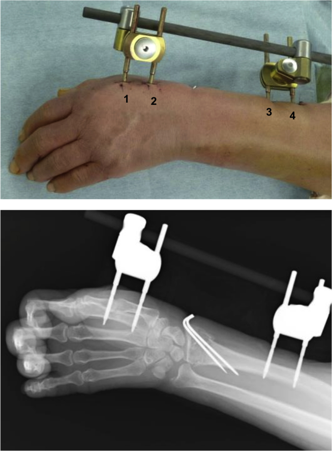
Methods: Unstable, displaced fractures of the distal radius that were medically suitable for external fixation were treated using external fixation pins coated and uncoated with an FGF-2-apatite composite layer. The coated pin group (n = 5) comprised 5 women (average age, 70.4 ± 5.9 years), whereas the uncoated pin group (n = 10) comprised 8 women and 2 men (average age, 64.4 ± 11.7 years). The average duration of external fixation was 40.8 ± 1.3 and 41.6 ± 2.1 days for the coated and uncoated pin groups, respectively.
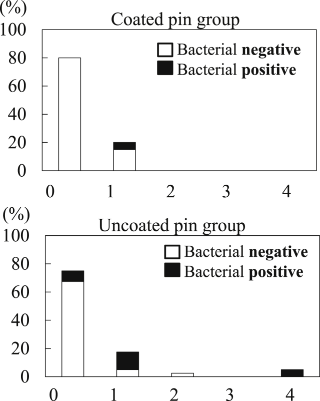
Results: All patients achieved fracture union. One patient in the uncoated group had severe pin tract infection on the day of pin extraction. No pin loosening or difficulty in pin removal was observed in either group. Bacterial growth was present in 5% and 25% of the pin sites in the coated and uncoated groups, respectively (p = 0.059). No adverse events such as tumor formation were observed for more than 2 years after surgery in the coated pin group.
Conclusions: This study clarified the safety and feasibility of using pins coated with an FGF-2-apatite composite layer for the external fixation of distal radius fractures.
Keywords: Apatite; Coating; Distal radius fractures; External fixation; Fibroblast growth factor 2 (FGF-2); Initial clinical trial; Safety.
Figures
Similar articles
-
Potential of Titanium Pins Coated with Fibroblast Growth Factor-2-Calcium Phosphate Composite Layers to Reduce the Risk of Impaired Bone-Pin Interface Strength in the External Fixation of Distal Radius Fractures.J Clin Med. 2024 May 22;13(11):3040. doi: 10.3390/jcm13113040. J Clin Med. 2024. PMID: 38892751 Free PMC article.
-
Reducing the risk of impaired bone apposition to titanium screws with the use of fibroblast growth factor-2-apatite composite layer coating.J Orthop Surg Res. 2017 Jan 5;12(1):1. doi: 10.1186/s13018-016-0501-z. J Orthop Surg Res. 2017. PMID: 28057033 Free PMC article.
-
Enhanced wound healing associated with Sharpey's fiber-like tissue formation around FGF-2-apatite composite layers on percutaneous titanium screws in rabbits.Arch Orthop Trauma Surg. 2012 Jan;132(1):113-21. doi: 10.1007/s00402-011-1381-7. Epub 2011 Sep 9. Arch Orthop Trauma Surg. 2012. PMID: 21904932
-
State of the art review: techniques to avoid pin loosening and infection in external fixation.J Orthop Trauma. 2002 Mar;16(3):189-95. doi: 10.1097/00005131-200203000-00009. J Orthop Trauma. 2002. PMID: 11880783 Review.
-
Hydroxyapatite-coated external fixation pins.Expert Rev Med Devices. 2005 Jul;2(4):465-71. doi: 10.1586/17434440.2.4.465. Expert Rev Med Devices. 2005. PMID: 16293085 Review.
Cited by
-
Inorganic Nanoparticles in Bone Healing Applications.Pharmaceutics. 2022 Mar 31;14(4):770. doi: 10.3390/pharmaceutics14040770. Pharmaceutics. 2022. PMID: 35456604 Free PMC article. Review.
-
Role of coatings and materials of external fixation pins on the rates of pin tract infection: A systematic review and meta-analysis.World J Orthop. 2021 Nov 18;12(11):920-930. doi: 10.5312/wjo.v12.i11.920. eCollection 2021 Nov 18. World J Orthop. 2021. PMID: 34888152 Free PMC article.
-
Do Stainless-Steel Pins Coated with Fibroblast Growth Factor-Calcium Phosphatase Composite Layers Have Anti-Infective Effects?Medicina (Kaunas). 2024 Aug 30;60(9):1419. doi: 10.3390/medicina60091419. Medicina (Kaunas). 2024. PMID: 39336460 Free PMC article.
-
Bioactive Coatings Based on Hydroxyapatite, Kanamycin, and Growth Factor for Biofilm Modulation.Antibiotics (Basel). 2021 Feb 5;10(2):160. doi: 10.3390/antibiotics10020160. Antibiotics (Basel). 2021. PMID: 33562515 Free PMC article.
-
Fixation of delayed distal radial fracture involving metaphyseal diaphyseal junction in adolescents: a comparative study of crossed Kirschner-wiring and non-bridging external fixator.BMC Musculoskelet Disord. 2020 Jun 9;21(1):365. doi: 10.1186/s12891-020-03404-0. BMC Musculoskelet Disord. 2020. PMID: 32517675 Free PMC article.
References
-
- W-Dahl A., Toksvig-Larsen S., Lindstrand A. No difference between daily and weekly pin site care: a randomized study of 50 patients with external fixation. Acta Orthop Scand. 2003;74:704–708. - PubMed
-
- Wassall M.A., Santin M., Isalberti C., Cannas M., Denyer S.P. Adhesion of bacteria to stainless steel and silver-coated orthopedic external fixation pins. J Biomed Mater Res. 1997;36:325–330. - PubMed
-
- Moroni A., Faldini C., Marchetti S., Manca M., Consoli V., Giannini S. Improvement of the bone-pin interface strength in osteoporotic bone with use of hydroxyapatite-coated tapered external-fixation pins. A prospective, randomized clinical study of wrist fractures. J Bone Joint Surg Am. 2001;83:717–721. - PubMed
-
- Nakamura H., Matsuno T., Hashimoto Y., Nakamura T., Mataga I. Comparison of a hydroxyapatite-coated and an anodic oxidized titanium implant for experimentally induced peri-implantitis: macroscopic and novel radiographic evaluations in a canine model. J Hard Tissue Biol. 2015;24:347–355.
LinkOut - more resources
Full Text Sources
Other Literature Sources
