. 2019 Jan;43(1):95-104.
doi: 10.1016/j.jgr.2017.09.004. Epub 2017 Oct 16.

Ginsenoside compound K protects human umbilical vein endothelial cells against oxidized low-density lipoprotein-induced injury via inhibition of nuclear factor-κB, p38, and JNK MAPK pathways

Affiliations

Ginsenoside compound K protects human umbilical vein endothelial cells against oxidized low-density lipoprotein-induced injury via inhibition of nuclear factor-κB, p38, and JNK MAPK pathways

Shan Lu et al. J Ginseng Res. 2019 Jan.

Erratum in

Abstract

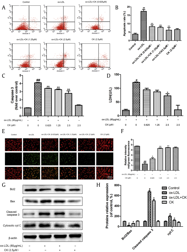
Background: Oxidized low-density lipoprotein (ox-LDL) causes vascular endothelial cell inflammatory response and apoptosis and plays an important role in the development and progression of atherosclerosis. Ginsenoside compound K (CK), a metabolite produced by the hydrolysis of ginsenoside Rb1, possesses strong anti-inflammatory effects. However, whether or not CK protects ox-LDL-damaged endothelial cells and the potential mechanisms have not been elucidated.

Methods: In our study, cell viability was tested using a 3-(4, 5-dimethylthiazol-2yl-)-2,5-diphenyl tetrazolium bromide (MTT) assay. Expression levels of interleukin-6, monocyte chemoattractant protein-1, tumor necrosis factor-α, intercellular adhesion molecule-1, and vascular cell adhesion molecule-1 were determined by enzyme-linked immunosorbent assay and Western blotting. Mitochondrial membrane potential (ΔΨm) was detected using JC-1. The cell apoptotic percentage was measured by the Annexin V/ propidium iodide (PI) assay, lactate dehydrogenase, and caspase-3 expression. Apoptosis-related proteins, nuclear factor (NF)-κB, and mitogen-activated protein kinases (MAPK) signaling pathways protein expression were quantified by Western blotting.

Results: Our results demonstrated that CK could ameliorate ox-LDL-induced human umbilical vein endothelial cells (HUVECs) inflammation and apoptosis, NF-κB nuclear translocation, and the phosphorylation of p38 and c-Jun N-terminal kinase (JNK). Moreover, anisomycin, an activator of p38 and JNK, significantly abolished the anti-apoptotic effects of CK.

Conclusion: These results demonstrate that CK prevents ox-LDL-induced HUVECs inflammation and apoptosis through inhibiting the NF-κB, p38, and JNK MAPK signaling pathways. Thus, CK is a candidate drug for atherosclerosis treatment.

Keywords: apoptosis; ginsenoside compound K; human umbilical vein endothelial cells; inflammation; oxidized low-density lipoprotein.


Figures

Fig. 1
Fig. 1
CK reduces ox-LDL-induced cytotoxicity. (A) Molecular structure of CK. (B) HUVECs were incubated with CK alone (0.625μM, 1.25μM, and 2.5 μM) for 12 h, and cell viability was assayed by the MTT assay. (C) HUVECs were pretreated with various concentrations of CK (0.625μM, 1.25μM, and 2.5μM) for 12 h and incubated with ox-LDL (80 μg/mL) for an additional 24 h. Then, cell viability was assessed by the MTT assay. Values are expressed as mean ± SD, n = 3. ##p < 0.01 versus control group; ∗∗p < 0.01 versus ox-LDL group. CK, compound K; HUVECs, human umbilical vein endothelial cells; MTT, 3-(4, 5-dimethylthiazol-2yl-)-2,5-diphenyl tetrazolium bromide; NS, no significance; ox-LDL, oxidized low-density lipoprotein; SD, standard deviation.
Fig. 2
Fig. 2
CK attenuates ox-LDL-induced HUVEC inflammation. (A–C) HUVECs were pretreated with various concentrations of CK (0.625μM, 1.25μM, and 2.5μM) for 12 h and incubated with or without ox-LDL (80 μg/mL) for an additional 24 h. The levels of IL-6, MCP-1 and TNF-α in the culture supernatant were assayed with enzyme-linked immunosorbent assay. (D) HUVECs were pretreated with CK (2.5μM) for 12 h, followed by treatment with ox-LDL (80 μg/mL) for another 24 h. VCAM-1, ICAM-1, and β-actin were evaluated by Western blot analysis. (E) Densitometric analysis was used to quantify the levels of VCAM-1 and ICAM-1. Values are expressed as mean ± SD, n = 3. #p < 0.05, ##p < 0.01 versus control group; p < 0.05, ∗∗p < 0.01 versus ox-LDL group. CK, compound K; HUVECs, human umbilical vein endothelial cells; ICAM-1, intercellular adhesion molecule-1; IL-6, interleukin-6; MCP-1, monocyte chemoattractant protein-1; ox-LDL, oxidized low-density lipoprotein; SD, standard deviation; TNF-α, tumor necrosis factor-α; VCAM-1, vascular cell adhesion molecule-1.
Fig. 3
Fig. 3
CK inhibits ox-LDL-induced HUVECs apoptosis. HUVECs were treated with ox-LDL (80μg/mL) in the presence or absence of various concentrations of CK (0.625μM, 1.25μM, and 2.5μM) for 12h, and incubated with or without ox-LDL (80 μg/mL) for additional 24 h. (A) A scatter diagram of apoptotic HUVECs was detected through annexin V/PI double staining by flow cytometry. (B) Quantity analysis of the percentages of apoptotic cells. (C) Caspase-3 activity was measured using a fluorometric assay. (D) The effect of CK on LDH level in HUVECs was measured using an LDH assay kit. (E) Cells were dyed with JC-1 and then detected using a fluorescence microscope. (F) Quantitative analysis of JC-1 red/green rates. (G) HUVECs were treated with ox-LDL (80μg/mL) in the presence or absence of CK (2.5μM) for 12h, and incubated with or without ox-LDL (80 μg/mL) for additional 24 h. Bcl-2, Bax, cleaved caspase-3, cyt C, and β-actin were evaluated by Western blot analysis. (H) Densitometric analysis was used to quantify the levels of bcl-2, bax, cleaved caspase-3, and cyt C. Values are expressed as the mean ± SD, n = 3. #p < 0.05, ##p < 0.01 ox-LDL group versus control group; p < 0.05, ∗∗p < 0.01 versus ox-LDL group. CK, compound K; HUVECs, human umbilical vein endothelial cells; LDH, lactate dehydrogenase; ox-LDL, oxidized low-density lipoprotein; SD, standard deviation; V/PI, Annexin V/ propidium iodide.
Fig. 4
Fig. 4
CK reduces ox-LDL-induced HUVECs inflammation through inhibiting the NF-κB pathway. (A) HUVECs were pretreated with CK (2.5μM) for 12 h, followed by treatment with ox-LDL (80 μg/mL) for another 24 h. NF-κB p65 immunoreactivity was observed by immunofluorescence assay. (B) HUVECs were treated as described in (A). LOX-1, p-IKKβ, p-IκBα, IκBα, NF-κB p65 (nuclear), NF-κB p65 (cytoplasm), NF-κB p65, Histone H3, and β-actin were evaluated by Western blot analysis. (C) Densitometric analysis was used to quantify the levels of LOX-1, p-IKKα/β, p-IκB and IκB. (D) Densitometric analysis was used to quantify the levels of NF-κB p65. Values are expressed as the mean ± SD, n = 3. #p < 0.05, ##p < 0.01 versus control group; p < 0.05, ∗∗p < 0.01 versus ox-LDL group. CK, compound K; DAPI, 4′,6-diamidino-2-phenylindole; HUVECs, human umbilical vein endothelial cells; LOX-1, lectin-like ox-LDL receptor-1; NF-κB, nuclear factor-κB; ox-LDL, oxidized low-density lipoprotein; SD, standard deviation.
Fig. 5
Fig. 5
CK prevents ox-LDL-induced HUVECs apoptosis through inhibiting the MAPK pathway. (A) HUVECs were pretreated with CK (2.5μM) for 12 h, followed by treatment with ox-LDL (80 μg/mL) for another 24 h. The expression levels of phosphorylated and total ERK1/2, p38, and JNK were detected by Western blot analysis. (B) Densitometric analysis was used to quantify the ratios of phospho-p38 to total p38, p-JNK to total JNK, p-ERK1/2 to total ERK1/2. (C) HUVECs were treated with CK (2.5μM) in the presence or absence of anisomycin (1μM) for 1 h, followed by treatment with ox-LDL (80 μg/mL) for another 24 h. Representative Western blot analysis of phosphorylated and total p38, and JNK in HUVECs was performed. (D) Densitometric analysis was used to quantify the levels of p-p38, p-JNK, and p-ERK1/2. Values are expressed as mean ± SD, n = 3. #p < 0.05, ##p < 0.01 versus control group; p < 0.05, ∗∗p < 0.01 versus ox-LDL group; $p < 0.05, $$p < 0.01 versus ox-LDL and CK treatment group. AM, anisomycin; CK, compound K; ERK1/2, extracellular signal-regulated protein kinases 1 and 2; HUVECs, human umbilical vein endothelial cells; JNK, c-Jun N-terminal kinase; MAPK, mitogen-activated protein kinase; NF-κB, nuclear factor-κB; ox-LDL, oxidized low-density lipoprotein; SD, standard deviation.
Fig. 6
Fig. 6
Anisomycin reverses the anti-apoptotic activity of CK. (A) HUVECs were treated with CK (2.5μM) in the presence or absence of anisomycin (1μM) for 1 h, followed by treatment with ox-LDL (80 μg/mL) for another 24 h. Cell viability was detected by the MTT assay. (B) HUVECs were treated as described in (A), and the expression levels of Bax, Bcl-2, cleaved caspase-3, and cyt C were detected by Western blot analysis. (C) Densitometric analysis was used to quantify the levels of Bax, Bcl-2, cleaved caspase-3, and cyt C. Values are expressed as the mean ± SD, n = 3. #p < 0.05, ##p < 0.01 versus control group; p < 0.05, ∗∗p < 0.01 versus ox-LDL group; $p < 0.05, $$p < 0.01 versus ox-LDL and CK treatment group. AM, anisomycin; CK, compound K; HUVECs, human umbilical vein endothelial cells; MTT, (4, 5-dimethylthiazol-2yl-)-2,5-diphenyl tetrazolium bromide; ox-LDL, oxidized low-density lipoprotein; SD, standard deviation.
Fig. 7
Fig. 7
Hypothetical mechanism by which CK prevents ox-LDL-induced HUVECs injury. CK protects ox-LDL-induced HUVECs injury through LOX-1-mediated NF-κB, p-38, and JNK MAPK pathways. CK, compound K; ERK1/2, extracellular signal-regulated protein kinases 1 and 2; HUVECs, human umbilical vein endothelial cells; ICAM-1, intercellular adhesion molecule-1; IL-6, interleukin-6; JNK, c-Jun N-terminal kinase; LOX-1, lectin-like ox-LDL receptor-1; MAPK, mitogen-activated protein kinase; MCP-1, monocyte chemoattractant protein-1; NF-κB, nuclear factor-κB; ox-LDL, oxidized low-density lipoprotein; SD, standard deviation; TNF-α, tumor necrosis factor-α; VCAM-1, vascular cell adhesion molecule-1.

References

    1. Siegel D., Devaraj S., Mitra A., Raychaudhuri S.P., Raychaudhuri S.K., Jialal I. Inflammation, atherosclerosis, and psoriasis. Clin Rev Allergy Immunol. 2013;44:194–204. - PubMed
    1. Gutierrez E., Flammer A.J., Lerman L.O., Elizaga J., Lerman A., Fernandez-Aviles F. Endothelial dysfunction over the course of coronary artery disease. Eur Heart J. 2013;34:3175–3181. - PMC - PubMed
    1. Matsuura E., Atzeni F., Sarzi-Puttini P., Turiel M., Lopez L.R., Nurmohamed M.T. Is atherosclerosis an autoimmune disease? BMC Med. 2014;12:47. - PMC - PubMed
    1. Chistiakov D.A., Orekhov A.N., Bobryshev Y.V. LOX-1-mediated effects on vascular cells in atherosclerosis. Cell Physiol Biochem. 2016;38:1851–1859. - PubMed
    1. Ogura S., Kakino A., Sato Y., Fujita Y., Iwamoto S., Otsui K., Yoshimoto R., Sawamura T. Lox-1: the multifunctional receptor underlying cardiovascular dysfunction. Circ J. 2009;73:1993–1999. - PubMed