

New Viewpoint of Surface Anatomy Using the Curved Sectional Planes of a Male Cadaver
- PMID: 30662383
- PMCID: PMC6335124
- DOI: 10.3346/jkms.2019.34.e15
New Viewpoint of Surface Anatomy Using the Curved Sectional Planes of a Male Cadaver
Abstract
Background: The curved sectional planes of the human body can provide a new approach of surface anatomy that the classical horizontal, coronal, and sagittal planes cannot do. The purpose of this study was to verify whether the curved sectional planes contribute to the morphological comprehension of anatomical structures.
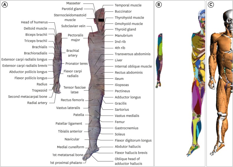
Methods: By stacking the sectioned images of a male cadaver, a volume model of the right half body was produced (voxel size 1 mm). The sectioned images with the segmentation data were also used to build another volume model. The volume models were peeled and rotated to be screen captured. The captured images were loaded on user-friendly browsing software that had been made in the laboratory.
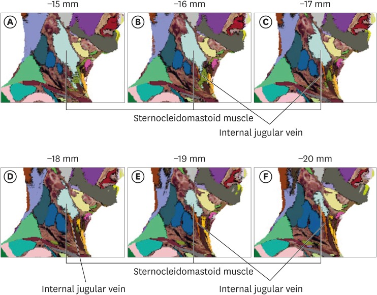
Results: The browsing software was downloadable from the authors' homepage (anatomy.co.kr). On the software, the volume model was peeled at 1 mm thicknesses and rotated at 30 degrees. Since the volume models were made from the cadaveric images, actual colors of the structures were displayed in high resolution. Thanks to the segmentation data, the structures on the volume model could be automatically annotated. Using the software, the sternocleidomastoid muscle and the internal jugular vein in the neck region, the cubital fossa in the upper limb region, and the femoral triangle in the lower limb region were observed to be described.
Conclusion: For the students learning various medical procedures, the software presents the needed graphic information of the human body. The curved sectional planes are expected to be a tool for disciplinary convergence of the sectional anatomy and surface anatomy.
Keywords: Cadaver; Cross-Sectional Anatomy; Education; Three-Dimensional Imaging; Visible Human Projects.
Conflict of interest statement
Disclosure: The authors have no potential conflicts of interest to disclose.
Figures


Comment in
-
Convergence by Breaking Stereotype.J Korean Med Sci. 2019 Jan 9;34(3):e26. doi: 10.3346/jkms.2019.34.e26. eCollection 2019 Jan 21. J Korean Med Sci. 2019. PMID: 30662390 Free PMC article. No abstract available.
References
-
- Donnelly L, Patten D, White P, Finn G. Virtual human dissector as a learning tool for studying cross-sectional anatomy. Med Teach. 2009;31(6):553–555. - PubMed
-
- Oh CS, Kim JY, Choe YH. Learning of cross-sectional anatomy using clay models. Anat Sci Educ. 2009;2(4):156–159. - PubMed
-
- Shin DS, Chung MS, Park HS, Park JS, Hwang SB. Browsing software of the Visible Korean data used for teaching sectional anatomy. Anat Sci Educ. 2011;4(6):327–332. - PubMed
-
- de Barros N, Rodrigues CJ, Rodrigues AJ, Jr, de Negri Germano MA, Cerri GG. The value of teaching sectional anatomy to improve CT scan interpretation. Clin Anat. 2001;14(1):36–41. - PubMed
MeSH terms
LinkOut - more resources
Full Text Sources
