Review
. 2019 Jan 4:9:3083.
doi: 10.3389/fimmu.2018.03083. eCollection 2018.

The C/EBP Homologous Protein (CHOP) Transcription Factor Functions in Endoplasmic Reticulum Stress-Induced Apoptosis and Microbial Infection

Affiliations
Review

The C/EBP Homologous Protein (CHOP) Transcription Factor Functions in Endoplasmic Reticulum Stress-Induced Apoptosis and Microbial Infection

Hai Hu et al. Front Immunol. .

Abstract

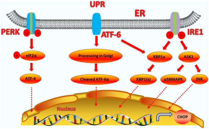
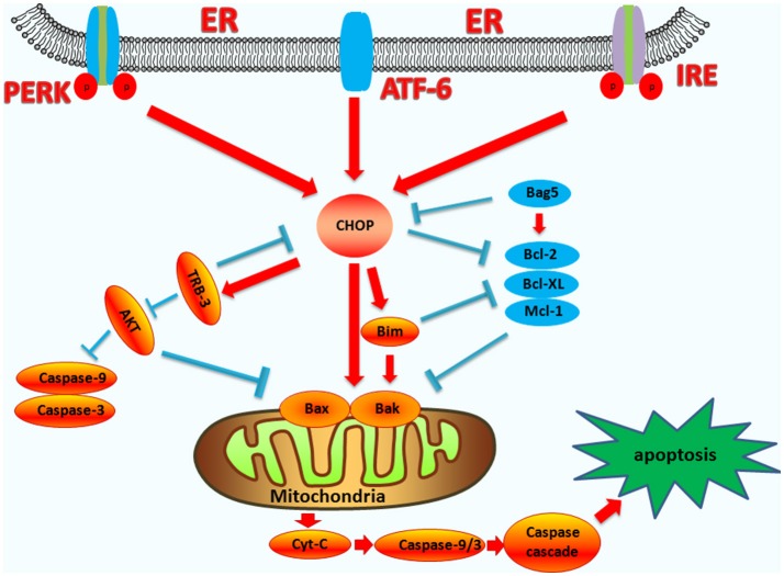
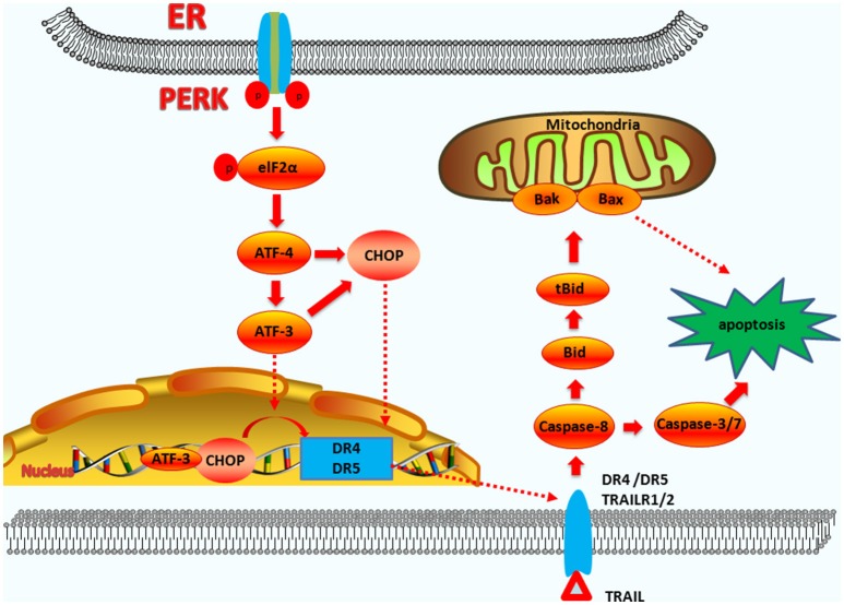
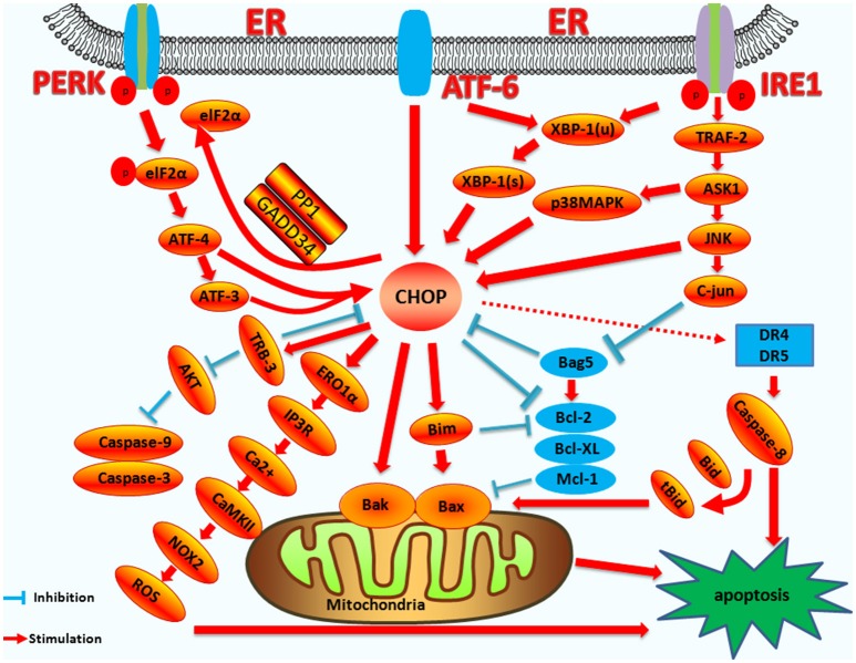
Apoptosis is a form of cell death by which the body maintains the homeostasis of the internal environment. Apoptosis is an initiative cell death process that is controlled by genes and is mainly divided into endogenous pathways (mitochondrial pathway), exogenous pathways (death receptor pathway), and apoptotic pathways induced by endoplasmic reticulum (ER) stress. The homeostasis imbalance in ER results in ER stress. Under specific conditions, ER stress can be beneficial to the body; however, if ER protein homeostasis is not restored, the prolonged activation of the unfolded protein response may initiate apoptotic cell death via the up-regulation of the C/EBP homologous protein (CHOP). CHOP plays an important role in ER stress-induced apoptosis and this review focuses on its multifunctional roles in that process, as well as its role in apoptosis during microbial infection. We summarize the upstream and downstream pathways of CHOP in ER stress induced apoptosis. We also focus on the newest discoveries in the functions of CHOP-induced apoptosis during microbial infection, including DNA and RNA viruses and some species of bacteria. Understanding how CHOP functions during microbial infection will assist with the development of antimicrobial therapies.

Keywords: C/EBP homologous protein; apoptosis; bacteria; endoplasmic reticulum stress; microorganisms; virus.


Figures

Figure 1
Figure 1
Upstream regulatory pathway of CHOP. The three signaling branches of UPR lead to CHOP transcription. Activated PERK phosphorylates eIF2α, which results in the translation of ATF4. Subsequently, ATF4 translocates to the nucleus, which increases the expression of CHOP and ATF3. CHOP and ATF3 then bind to the promoters downstream genes. ATF6 translocates to Golgi apparatus, where it is activated by proteolysis. Activated ATF6 transcriptionally upregulates CHOP expression. Additionally, ATF6 can regulate XBP-1 to activate CHOP. On one hand, the activation of IRE1α processes unspliced XBP1 mRNA to create activated XBP1(s), which enters the nucleus and controls the expression of CHOP. On the other hand, IRE1α activates apoptotic signaling kinase 1 (ASK1), which in turn phosphorylates p38MAPK and JNK to activate CHOP.
Figure 2
Figure 2
The functions of CHOP in endogenous-pathway-induced apoptosis. CHOP triggers the intrinsic apoptotic pathway through the inhibition of BCL-2, BCL-XL, MCL-1, and the upregulation of BIM, which regulates BAX-BAK-mediated mitochondrial outer membrane permeabilization. This leads to cytochrome c release and the caspase cascade. Bag5 can reduce CHOP expression and increase Bcl-2 gene expression. CHOP can also regulate the apoptosis of cells by up-regulating the expression of the TRB3 gene, preventing Akt phosphorylation, which inhibits the activity of caspases-3/9.
Figure 3
Figure 3
The functions of CHOP in exogenous-pathway-induced apoptosis. CHOP triggers the extrinsic apoptotic pathway through the upregulation of DR4 and DR5. PERK can induce ATF4 expression, which results in CHOP and ATF3 expression. CHOP and ATF3 then bind to the promoters of the DR4 and DR5 genes, upregulating their expression. TRAIL combining with DR4 and DR5 regulates the caspase 8-mediated cascade, which activates caspase3/7 to lead apoptosis. On the other hand, Bid is cleavaged into tBid. tBid then regulates the BAX-BAK-mediated mitochondrial apoptosis pathways.
Figure 4
Figure 4
The mechanism of CHOP regulation in ER stress induced apoptosis. Upstream regulatory pathway: Activated PERK phosphorylates eIF2α, which results in the translation of ATF4. ATF4 translocates to the nucleus, which increases the expression of CHOP and ATF3. CHOP and ATF3 then bind to the promoters of the target genes, upregulating their expression. ATF6 translocates to Golgi apparatus where it is activated by proteolysis. Activated ATF6 transcriptionally upregulates CHOP expression. Additionally, ATF6 can regulate XBP-1 to activate CHOP. Activation of IRE1α processes unspliced XBP-1 mRNA into activated XBP1(s), which enters the nucleus and controls the expression of CHOP. IRE1α activates the apoptotic signaling kinase 1 (ASK1), which in turn phosphorylates p38 MAPK/JNK, and activates CHOP. In addition, Bag5 can reduce CHOP expression and increase Bcl-2 gene expression. Downstream regulatory pathway: CHOP triggers the intrinsic apoptotic pathway through the inhibition of BCL-2, BCL-XL, MCL-1, and the upregulation of BIM, which regulates BAX-BAK-mediated mitochondrial outer membrane permeabilization. This leads to cytochrome c release and the caspase cascade. CHOP can also regulate apoptosis by upregulating the expression of the TRB3 gene and preventing Akt phosphorylation, which inhibits the activity of caspases-3/9. CHOP triggers the extrinsic apoptotic pathway through the upregulation of DR4 and DR5, which regulate the caspase-8-mediated cascade. This leads to Bid cleavage into tBid, which regulates the BAX-BAK-mediated mitochondrial apoptosis pathways. CHOP can also trigger the ERO1α-IP3R-Ca2+-CaMKII pathway. ROS can also trigger Ca2+-dependent mitochondrial apoptosis. CHOP can directly activate GADD34 (DNA damage protein), which, combined with phosphatase 1 protein (PP1), dephosphorylates eIF2α, and results in protein translation recovery, increased ER stress, and cell apoptosis.

References

    1. Sano R, Reed JC. ER stress-induced cell death mechanisms. Biochim Biophys Acta (2013) 1833:3460–70. 10.1016/j.bbamcr.2013.06.028 - DOI - PMC - PubMed
    1. Han J, Kaufman RJ. The role of ER stress in lipid metabolism and lipotoxicity. J Lipid Res. (2016) 57:1329–38. 10.1194/jlr.R067595 - DOI - PMC - PubMed
    1. Sovolyova N, Healy S, Samali A, Logue SE. Stressed to death - mechanisms of ER stress-induced cell death. Biol Chem. (2014) 395:1–13. 10.1515/hsz-2013-0174 - DOI - PubMed
    1. Luethy JD, Holbrook NJ. Activation of the gadd153 promoter by genotoxic agents: a rapid and specific response to DNA damage. Cancer Res. (1992) 52:5–10. - PubMed
    1. Pahl HL. Signal transduction from the endoplasmic reticulum to the cell nucleus. Physiol Rev. (1999) 79:683–701. 10.1152/physrev.1999.79.3.683 - DOI - PubMed

Publication types

MeSH terms

Substances