. 2018 Nov 15;10(11):3650-3663.
eCollection 2018.

Vincamine exerts protective effect on spiral ganglion neurons in endolymphatic hydrops guinea pig models

Affiliations

Vincamine exerts protective effect on spiral ganglion neurons in endolymphatic hydrops guinea pig models

Yi Li et al. Am J Transl Res. .

Abstract

The aim of this study was to investigate the protective effects of vincamine in endolymphatic hydrops (ELH). After ELH guinea pigs treated by vincamine, the concentration of VAP in plasma, and the levels of cAMP, MDA, SOD, GSH-Px in right cochlea were measured using spectrophotometric method. The V2R, NMDAR1, p-NMDAR1, AQP2, p-AQP2, caspase3/9 and c-caspase3/9 expressions in right cochlea were detected using western blot analysis. The cochlear hydrops degree and SGNs density were evaluated by hemotoxylin and eosin staining (HE) test. Normal hearing and vestibular function were warranted by the tests of auditory brainstem response (ABR) and electronystagmography (ENG). After glutamate-injured SGNs treated with vincamine, the MDA, SOD GSH-Px, NGF, BDNF, NT3, NT4 and Trks levels were measured. Meanwhile, the Bcl2, Bax, NMDAR1, p-NMDAR1, PI3K, p-PI3K, Akt, p-Akt, caspase3/9 and cleaved-caspase3/9 expression levels were detected. Furthermore, the viability, apoptosis and necrosis of SGNs were tested by MTT and Hoechst/PI staining methods. The results indicated that vincamine could significantly inhibit the expression levels of cAMP, MDA, V2R, p-NMDAR1, p-AQP2 and c-caspase-3/-9 in cochlea, alleviate the cochlear hydrops degree, regulate the audiological and vestibular dysfunctions. The SGNs density, SOD and GSH-Px levels were also increased by vincamine. In vincamine-treating groups, the MDA, Bax, p-NMDAR1, and c-caspase3/9 levels were observably decreased, while SGNs survival, SOD, GSH, NGF, BDNF, NT3, NT4, Trks, Bcl2, p-PI3K, p-Akt expressions were improved. The present study indicated a novel use of vincamine in suppressing ELH formation by down-regulating the VAP/AQP2 signaling pathway. It also manifested that vincamine exerted protective effects on hearing via improving neurotrophin-dependent PI3K/Akt signaling pathway in SGNs.

Keywords: Meniere’s disease; PI3K/Akt; VAP/AQP2; endolymphatic hydrops; vincamine.


Conflict of interest statement

None.

Figures

Figure 1
Figure 1
The chemical structure of vincamine. Molecular formula: C21H26N2O3. Molecular weight: 354.44 g/mol.
Figure 2
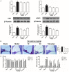
Figure 2
Vincamine down-regulated the cAMP, V2R, AQP2 levels, reduced the cochlear hydrops degree, improved spontaneous nystagmus and auditory brainstem response in ELH guinea pigs. ELH guinea pigs were treated with saline and/or vincamine (10 or 30 mg/kg/d) and/or tolvaptan (10 mg/kg/d) for 6 weeks. A. Plasma-AVP, cochlea-cAMP contents were measured using commercial assay kits, while cochlea-V2R, cochlea-AQP2 expression levels were analyzed via western blot with β-Tubulin as an invariant control for equal loading. Representative blots are shown with densitometry. Original full-size blots are presented in Figure S1. B. Representative examples of right cochlea from each experimental condition after treatment. Histological analysis confirmed the changes in endolymphatic space. The hydrops quantification was counted according to the ratio of SM area to SM + SV area (HE staining). C. Spontaneous nystagmus and auditory brainstem response were tested before and 6, 12 weeks after the surgery. Data are expressed as mean ± S.D. #P < 0.05, ##P < 0.01 vs sham; **P < 0.01, *P < 0.05 vs. model.
Figure 3
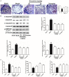
Figure 3
Vincamine inhibited the degeneration of SGNs, eliminated the accumulation of p-NMDAR1, c-caspase3/9 and MDA, and induced the secretion of SOD, GSH-Px in ight cochlea of ELH guinea pigs. ELH guinea pigs were treated with saline and/or vincamine (10 or 30 mg/kg/d) and/or tolvaptan (10 mg/kg/d) for 6 weeks. A. Observation and quantification of spiral ganglion neurons density in right cochlea. B. Related protein extractions of cochlear tissue were analyzed via western blot with β-Tubulin as an invariant control for equal loading. Representative blots are shown with densitometry. Original full-size blots are presented in Figure S2. C. MDA, SOD and GSH-Px contents were measured using commercial assay kits. Data are expressed as mean ± S.D. ##P < 0.01 vs sham; **P < 0.01, *P < 0.05 vs. model.
Figure 4
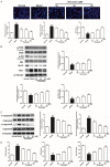
Figure 4
Vincamine prevented cell apoptosis and necrosis, improved cell survival, up-regulated p-PI3K, p-Akt and Bcl-2/Bax levels, eliminated c-caspase3/9 and MDA accumulation, and induced SOD, GSH-Px secretion in glutamate-injuried SGNs. SGNs isolated from embryonic rats were pretreated with vincamine (20-80 μM) for 1 h, and then treated with 2 mM glutamate for 23 h. A. SGNs apoptosis, necrosis and viability were measured using hoechst/PI dual-staining and MTT methods, respectively. B, C. Cell lysates were subjected to western blotting test and probed for the expression levels of PI3K, p-PI3K, Akt, p-Akt, caspase-3, cleaved-caspase3, caspase-9, cleaved-caspase9, Bcl-2 and Bax and β-Tubulin as the loading control. Representative blots are shown with densitometry. Original full-size blots are presented in Figure S3. D. MDA, SOD and CSH-Px levels in SGNs were tested according to the commercial assay kits. Data are expressed as mean ± S.D. #P < 0.05, ##P < 0.01 vs normal; **P < 0.01, *P < 0.05 vs. model.
Figure 5
Figure 5
Vincamine increased NGF, BDNF, NT3, NT4 and Trks expression levels, and induce fluorescence expression in glutamate-injuried SGNs. SGNs isolated from embryonic rats were pretreated with vincamine (20-80 μM) for 1 h, and then treated with 2 mM glutamate for 23 h. A. Immunofluorescence technique was used measuring the total expression of TrkA+ TrkB+ TrkC. B. Cell lysates were subjected to ELISA method and probed for the expression levels of NGF, BDNF, NT3 and NT4. Data are expressed as mean ± S.D. #P < 0.05, ##P < 0.01 vs normal; **P < 0.01, *P < 0.05 vs. model.

Similar articles

Cited by

References

    1. Lopez-Escamez JA, Carey J, Chung WH, Goebel JA, Magnusson M, Mandalà M, Newman-Toker DE, Strupp M, Suzuki M, Trabalzini F, Bisdorff A Classification Committee of the Barany Society; Japan Society for Equilibrium Research; European Academy of Otology and Neurotology (EAONO); Equilibrium Committee of the American Academy of Otolaryngology-Head and Neck Surgery (AAO-HNS); Korean Balance Society. Diagnostic criteria for Meniere’s disease. J Vestib Res. 2015;25:1–7. - PubMed
    1. Thai-Van H, Bounaix MJ, Fraysse B. Meniere’s disease: pathophysiology and treatment. Drugs. 2001;61:1089–1102. - PubMed
    1. Maekawa C, Kitahara T, Kizawa K, Okazaki S, Kamakura T, Horii A, Imai T, Doi K, Inohara H, Kiyama H. Expression and translocation of aquaporin-2 in the endolymphatic sac in patients with Meniere’s disease. J Neuroendocrinol. 2010;22:1157–1164. - PubMed
    1. Naganuma H, Kawahara K, Tokumasu K, Satoh R, Okamoto M. Effects of arginine vasopressin on auditory brainstem response and cochlear morphology in rats. Auris Nasus Larynx. 2014;41:249–254. - PubMed
    1. Foster CA, Breeze RE. Endolymphatic hydrops in Meniere’s disease: cause, consequence, or epiphenomenon? Otol Neurotol. 2013;34:1210–1214. - PubMed

LinkOut - more resources