

Novel cancer cell lines derived from primary breast tumors in Chinese patients
- PMID: 30662642
- PMCID: PMC6325518
Novel cancer cell lines derived from primary breast tumors in Chinese patients
Abstract
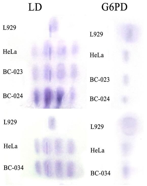
Although many breast cancer cell lines have been used for cancer research during the past several decades, few have originated from primary tumors in Asian patients. Moreover, the incidence of breast cancer has been increasing rapidly in China during this time period. Therefore, it is essential to establish breast cancer cell lines from Chinese patients. Here, we report the establishment of three new breast cancer cell lines, designated BC-023, BC-024, and BC-034, from breast carcinoma tissues of three Chinese patients. These breast cancer cell lines grew as adherent monolayers with characteristic epithelial morphology and were maintained continuously in vitro with stable growth rates for at least 20 passages. No bacterial, fungal, or mycoplasma contamination was detected in any of the three cell lines. Additionally, these cells were human epidermal growth factor receptor 2 (C-erbB-2)-positive. All three cell lines had comparable population doubling times of 33-39 h and reproducibly formed colonies in soft agar. Furthermore, these cells displayed aggressive tumorigenicity. Thus, every characteristic of each of these cell lines meets the quality control standards of the American Type Culture Collection (ATCC). We used drug sensitivity testing with growth-inhibition assays and showed that these three lines expressed a wide range of sensitivities to cisplatin (DDP) and adriamycin (ADR). These studies indicate that these three novel cell lines may provide new models for studying cancer biology and for screening new drugs for breast cancer treatment, especially for the Chinese population.
Keywords: Breast cancer; cell lines; drug sensitivity.
Conflict of interest statement
None.
Figures


Similar articles
-
Establishment and characterization of three new human breast cancer cell lines derived from Chinese breast cancer tissues.Cancer Cell Int. 2009 Jan 2;9:2. doi: 10.1186/1475-2867-9-2. Cancer Cell Int. 2009. PMID: 19121212 Free PMC article.
-
Establishment and characterization of three stable Basal/HER2-positive breast cancer cell lines derived from Chinese breast carcinoma with identical missense mutations in the DNA-binding domain of TP53.Cancer Cell Int. 2018 Aug 17;18:118. doi: 10.1186/s12935-018-0617-9. eCollection 2018. Cancer Cell Int. 2018. PMID: 30140169 Free PMC article.
-
Establishment and characterisation of new cell lines from human breast tumours initially established as tumour xenografts in NMRI nude mice.Breast Cancer Res Treat. 1997 May;43(3):247-58. doi: 10.1023/a:1005756632293. Breast Cancer Res Treat. 1997. PMID: 9150904
-
The significance of heregulin in breast cancer tumor progression and drug resistance.Breast Cancer Res Treat. 1996;38(1):57-66. doi: 10.1007/BF01803784. Breast Cancer Res Treat. 1996. PMID: 8825123 Review.
-
Feasibility and barriers to rapid establishment of patient-derived primary osteosarcoma cell lines in clinical management.iScience. 2024 Jun 19;27(9):110251. doi: 10.1016/j.isci.2024.110251. eCollection 2024 Sep 20. iScience. 2024. PMID: 39286504 Free PMC article. Review.
Cited by
-
miR‑125a‑5p reverses epithelial‑mesenchymal transition and restores drug sensitivity by negatively regulating TAFAZZIN signaling in breast cancer.Mol Med Rep. 2021 Nov;24(5):812. doi: 10.3892/mmr.2021.12452. Epub 2021 Sep 22. Mol Med Rep. 2021. PMID: 34549308 Free PMC article.
References
-
- Bertero C, Chamberlain Wilmoth M. Breast cancer diagnosis and its treatment affecting the self: a meta-synthesis. Cancer Nurs. 2007;30:194–202. quiz 203-194. - PubMed
-
- Wiseman MJ. The world cancer research fund expert report: the next steps. IARC Sci Publ. 2002;156:523–524. - PubMed
-
- Soule HD, Vazguez J, Long A, Albert S, Brennan M. A human cell line from a pleural effusion derived from a breast carcinoma. J Natl Cancer Inst. 1973;51:1409–1416. - PubMed
-
- Engel LW, Young NA, Tralka TS, Lippman ME, O’Brien SJ, Joyce MJ. Establishment and characterization of three new continuous cell lines derived from human breast carcinomas. Cancer Res. 1978;38:3352–3364. - PubMed
-
- Shen C, Gu M, Song C, Miao L, Hu L, Liang D, Zheng C. The tumorigenicity diversification in human embryonic kidney 293 cell line cultured in vitro. Biologicals. 2008;36:263–268. - PubMed
LinkOut - more resources
Full Text Sources
Research Materials
Miscellaneous