. 2018 Oct 1;3(10):526-540.
doi: 10.1302/2058-5241.3.170049. eCollection 2018 Oct.

Management of supracondylar fractures of the humerus in children

Affiliations

Management of supracondylar fractures of the humerus in children

Alfonso Vaquero-Picado et al. EFORT Open Rev. .

Abstract

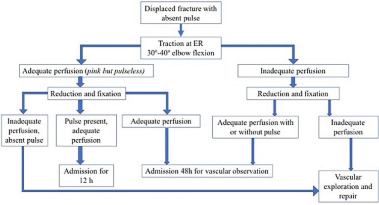
Supracondylar fractures of the humerus are the most frequent fractures of the paediatric elbow, with a peak incidence at the ages of five to eight years.Extension-type fractures represent 97% to 99% of cases. Posteromedial displacement of the distal fragment is the most frequent; however, the radial and median nerves are equally affected. Flexion-type fractures are more commonly associated with ulnar nerve injuries.Concomitant upper-limb fractures should always be excluded. To manage the vascular status, distal pulse and hand perfusion should be monitored. Compartment syndrome should always be borne in mind, especially when skin puckering, severe ecchymosis/swelling, vascular alterations or concomitant forearm fractures are present.Gartland's classification shows high intra- and inter-observer reliability. Type I is treated with casting. Surgical treatment is the standard for almost all displaced fractures. Type IV fractures can only be diagnosed intra-operatively.Closed reduction and percutaneous pinning is the gold standard surgical treatment. Open reduction via the anterior approach is indicated for open fractures, absence of the distal vascular flow for > 10 to 15 minutes after closed reduction, and failed closed reduction.Lateral entry pins provide stable fixation, avoiding the risk of iatrogenic ulnar nerve injury.About 10% to 20% of displaced supracondylar fractures present with alterations in vascular status. In most cases, fracture reduction restores perfusion.Neural injuries occur in 6.5% to 19% of cases involving displaced fractures. Most of them are neurapraxias and it is not routinely indicated to explore the nerve surgically. Cite this article: EFORT Open Rev 2018;3:526-540. DOI: 10.1302/2058-5241.3.170049.

Keywords: children; management; supracondylar fractures.


Conflict of interest statement

ICMJE Conflict of interest statement: None declared.

Figures

Fig. 1
Fig. 1
a) Skin puckering at the antecubital fossa should warn of a high energy fracture which transects the brachialis muscle and biceps. b) Very displaced type III fracture. When the fracture is very displaced, ‘S-deformity’ and skin puckering are usually present, and the possibility of neurovascular injury and compartment syndrome should be considered; c) The so-called ‘S-deformity’ is present in very displaced extension-type fractures.
Fig. 2
Fig. 2
a) The anterior humeral line (AHL) should traverse the middle third of the capitellum nucleus in normal anatomy and type I fractures; b, c) in extension-type fractures, the line crosses anteriorly.
Fig. 3
Fig. 3
Posterior fat-pad sign (asterisk) is specific to an occult elbow fracture in almost 75% of patients. However, anterior fat-pad elevation (arrow) is not so specific to fracture diagnosis and can appear in a normal elbow.
Fig. 4
Fig. 4
a) Baumann’s angle; b) ulnohumeral angle.
Fig. 5
Fig. 5
Gartland’s classification for extension fractures: a) type I; b) type II; c) type III.
Fig. 6
Fig. 6
Reduction technique. Reduction is performed under fluoroscopy. First, traction is applied. Then, coronal plane deformity is corrected, applying varus/valgus. Sagittal plane correction is then performed. The last step is fixation with smooth 1.8 to 2.0 mm K-wires.
Fig. 7
Fig. 7
When posteromedial displacement is present, forearm pronation helps with fracture reduction. Forearm supination will be a difficult reduction in these cases.
Fig. 8
Fig. 8
In very unstable fractures (such as type IV), a provisional pin in the distal fragment is helpful to achieve reduction. Once reduction is achieved, fixation is performed. In very unstable fractures, three-wire fixation is preferred.
Fig. 9
Fig. 9
Fracture fixated with crossed pins percutaneously. Ulnar nerve was traversed by medial pin and post-operative palsy was present.
Fig. 10
Fig. 10
According to Sankar et al, a type A error was defined as the failure to engage both fragments using two pins or more; a type B error was defined as the failure to achieve bicortical fixation with two pins or more; and a type C error was defined as inadequate pin spread to control rotation.
Fig. 11
Fig. 11
After reduction and fixation, the elbow should be immobilized at approximately 70° of flexion. Higher flexion not only will not improve results, as the reduction is maintained by pins, but also increases the risk of compartment syndrome.
Fig. 12
Fig. 12
Algorithm for the management of vascular injury in the context of a supracondylar fracture.
Fig. 13
Fig. 13
Fracture malunion in extension leads to a lack of flexion. It is an important point that stiffness in supracondylar fractures in children is not frequent and lack of mobility usually reflects a malunion.
Fig. 14
Fig. 14
Cubitus varus deformity as a result of malunion. Internal rotation of the distal fragment is the key problem in this deformity.
Fig. 15
Fig. 15
Ulnar nerve palsy is one of the complications related to cubitus varus deformity. In the figure, anterior transposition of the nerve is performed before the osteotomy for correction the deformity.

References

    1. Beaty JHKJ. Supracondylar fractures of the distal humerus. In: Beaty JH, Kasser JR, ed. Rockwood and Wilkins’ fractures in children. 6th ed. Philadelphia: Lippincott Williams and Wilkins; 2006:543-589.
    1. Omid R, Choi PD, Skaggs DL. Supracondylar humeral fractures in children. J Bone Joint Surg [Am] 2008;90-A(5):1121-1132. - PubMed
    1. Mulpuri K, Hosalkar H, Howard A. AAOS clinical practice guideline: the treatment of pediatric supracondylar humerus fractures. J Am Acad Orthop Surg 2012;20(5):328-330. - PubMed
    1. Cheng JC, Lam TP, Maffulli N. Epidemiological features of supracondylar fractures of the humerus in Chinese children. J Pediatr Orthop B 2001;10(1):63-67. - PubMed
    1. Moraleda L, Diez J, Valencia M. Is timing for paediatric supracondylar fractures related to pinning errors or complications? Podium presentation, POSNA Annual Meeting 2015, Atlanta, US.