Review
. 2019 Apr;38(4):523-527.
doi: 10.1097/ICO.0000000000001852.

Feasibility of Intraocular Projection for Treatment of Intractable Corneal Opacity

Affiliations
Review

Feasibility of Intraocular Projection for Treatment of Intractable Corneal Opacity

Sarah Y Shim et al. Cornea. 2019 Apr.

Abstract

Despite many decades of research and development, corneal opacity remains a leading cause of reversible blindness worldwide. Corneal transplantation and keratoprosthesis can restore corneal clarity, but both have well-known limitations. High-resolution electronic microdisplays may offer an alternative to traditional methods of treating corneal disease using an intraocular implant to project imagery onto the retina, obviating the need for a clear cornea. In this study, we review previous work and recent technologic developments relevant to the development of such an intraocular projection system.


Conflict of interest statement

The authors have no funding or conflicts of interest to disclose.

Figures

FIGURE 1.
FIGURE 1.
Intraocular projector. A unit implanted behind the ear receives power and video stream data from a radiofrequency transmitter. It delivers power and data to a projector implanted within the eye through a subcutaneous cable. Images on the display are then projected onto the retina, thereby bypassing the opacity of the cornea.
FIGURE 2.
FIGURE 2.
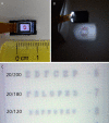
A, Commercially available microdisplay (Kopin) of size 7 × 4 mm, with a pixel pitch of 11 μm, and total resolution of 640 × 480 pixels. B, Image projected from a microdisplay using a projector of a size that can fit within the eye. C, The calculated visual acuity of this system is ∼20/130, which is demonstrated here by projection of an eye chart onto a camera sensor. Contrast can be improved with an optimized lens.
FIGURE 3.
FIGURE 3.
Schematic of a display-based visual prosthesis system. In the external portion, video captured by the camera is encoded by a processor. This signal, along with power, is sent through a radiofrequency transmission coil. In the implanted portion, a receiver unit separates the power and data signals and decodes the visual information to operate the microdisplay.
FIGURE 4.
FIGURE 4.
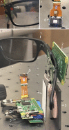
Prototype of the intraocular display electronics. A, External camera (1). B, Microdisplay (6). C, A camera (1) captures video, which is encoded by a processor (2), which sends the data and power through a transmission coil (3) to a receiver coil (4). The signal is then decoded by another processor (5), and the recovered video is displayed by the microdisplay (6).
FIGURE 5.
FIGURE 5.
Two methods of implantation. A–C, If the eye has had previous lensectomy and vitrectomy, a pars plana incision can be used. D and E, Alternatively, temporary keratoprosthesis can be used to perform lensectomy and vitrectomy. The projector can be inserted through the trephination incision, and the patient's original corneal button can be sutured back to close the wound. In both cases, the device haptics are externalized through small sclerotomies and fixated to the sclera.

References

    1. Gain P, Jullienne R, He Z, et al. Global survey of corneal transplantation and eye banking. JAMA Ophthalmol. 2016;134:167–173. - PubMed
    1. Bartels MC, Doxiadis II, Colen TP, et al. Long-term outcome in high-risk corneal transplantation and the influence of HLA-A and HLA-B matching. Cornea. 2003;22:552–556. - PubMed
    1. Saeed HN, Shanbhag S, Chodosh J. The Boston keratoprosthesis. Curr Opin Ophthalmol. 2017;28:390–396. - PubMed
    1. Lee WB, Shtein RM, Kaufman SC, et al. Boston keratoprosthesis: outcomes and complications: a report by the American Academy of Ophthalmology. Ophthalmology. 2015;122:1504–1511. - PubMed
    1. Iyer G, Srinivasan B, Agarwal S, et al. Boston type 2 keratoprosthesis-mid term outcomes from a tertiary eye care centre in India. Ocul Surf. 2018. (ePub ahead of print). - PubMed