. 2019 Mar;43(3):1289-1298.
doi: 10.3892/ijmm.2019.4063. Epub 2019 Jan 14.

Laminar shear stress alters endothelial KCa2.3 expression in H9c2 cells partially via regulating the PI3K/Akt/p300 axis

Affiliations

Laminar shear stress alters endothelial KCa2.3 expression in H9c2 cells partially via regulating the PI3K/Akt/p300 axis

Guojian Li et al. Int J Mol Med. 2019 Mar.

Abstract

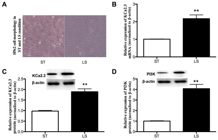
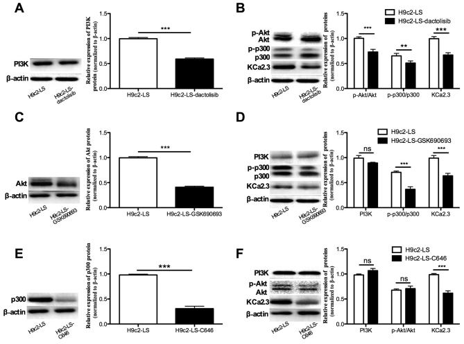
In cardiac tissues, myoblast atrial myocytes continue to be exposed to mechanical forces including shear stress. However, little is known about the effects of shear stress on atrial myocytes, particularly on ion channel function, in association with disease. The present study demonstrated that the Ca2+‑activated K+ channel (KCa)2.3 serves a vital role in regulating arterial tone. As increased intracellular Ca2+ levels and activation of histone acetyltransferase p300 (p300) are early responses to laminar shear stress (LSS) that result in the transcriptional activation of genes, the role of p300 and the phosphoinositide3‑kinase (PI3K)/protein kinase B (Akt) pathway, an intracellular pathway that promotes the growth and proliferation rather than the differentiation of adult cells, in the LSS‑dependent regulation of KCa2.3 in cardiac myoblasts was examined. In cultured H9c2 cells, exposure to LSS (15 dyn/cm2) for 12 h markedly increased KCa2.3 mRNA expression. Inhibiting PI3K attenuated the LSS‑induced increases in the expression and channel activity of KCa2.3, and decreased the phosphorylation levels of p300. The upregulation of these channels was abolished by the inhibition of Akt through decreasing p300 phosphorylation. ChIP assays indicated that p300 was recruited to the promoter region of the KCa2.3 gene. Therefore, the PI3K/Akt/p300 axis serves a crucial role in the LSS‑dependent induction of KCa2.3 expression, by regulating cardiac myoblast function and adaptation to hemodynamic changes. The key novel insights gained from the present study are: i) KCa2.3 was upregulated in patients with atrial fibrillation (AF) and in patients with AF combined with mitral value disease; ii) LSS induced a profound upregulation of KCa2.3 mRNA and protein expression in H9c2 cells; iii) PI3K activation was associated with LSS‑induced upregulation of the KCa2.3 channel; iv) PI3K activation was mediated by PI3K/Akt‑dependent Akt activation; and v) LSS induction of KCa2.3 involved the binding of p300 to transcription factors in the promoter region of the KCa2.3 gene.


Figures

Figure 1
Figure 1
Histological analysis of atrial interstitial fibrosis. Representative Masson trichrome staining of left atrial myocardium in the (A) SR, (B) AF and (C) AF combined with MVD groups (magnification, ×200; scale bar, 200 µm). (D) Percentage of areas of interstitial fibrous tissue in the three groups of patients; the bars indicate the standard deviation. **P<0.01 and ***P<0.001 vs. SR group. SR, sinus rhythm; AF, atrial fibrillation; MVD, mitral valve disease.
Figure 2
Figure 2
KCa2.3 gene expression analysis in clinical samples. (A) 2.66- and 3.1-fold increases in KCa2.3 expression levels were observed in the AF (n = 8) and AF combined with MVD (n=6) groups compared to the SR groups (n=6; P=0.006 and 0.005, respectively). (B) Increases in 85% and 63% in KCa2.3 expression was observed in the AF (n=8) and AF combined with MVD (n=6) groups compared to the SR group (n=6; P<0.001 and P=0.004, respectively). (C) Expression levels of KCa2.3 in SR, AF and AF combined with MVD groups were examined by immunofluorescence. Fluorescence was observed by laser-scanning microscopy. Scale bars, 50 µm. (D) Western blot analysis was performed to detect the protein expression of PI3K in patients with SR, AF and AF combined with MVD; β-actin was used as an internal reference protein. Densitometric quantification of the western blot analysis results are presented; the bars represents the mean ± standard deviation of three independent experiments. *P<0.05, **P<0.01 and ***P<0.001 vs. SR group. KCa, Ca2+-activated K+ channels; SR, sinus rhythm; AF, atrial fibrillation; MVD, mitral valve disease.
Figure 3
Figure 3
LS increases KCa2.3 partially via regulating PI3K expression. The effect of shear stress on the expression of KCa2.3 in H9c2 cells was determined by reverse transcription quantitative polymerase chain reaction and western blot analysis. (A) Exposure to LS (15 dynes/cm2) for 12 h markedly changed the morphology of the cells. (B) KCa2.3 mRNA levels were increased by 1.16-fold following LS. (C) KCa2.3 protein expression was increased by 0.91-fold following LS. (D) Exposure to LS (15 dynes/cm2) for 12 h upregulated the PI3K protein expression by 3.07-fold compared with the ST condition (P=0.0016). **P<0.01 vs. ST group. KCa, Ca2+-activated K+ channels; PI3K, phosphoinositide 3-kinase; Akt, protein kinase B; LS, laminar shear stress condition; ST, static culture conditions.
Figure 4
Figure 4
Activation of the PI3K/Akt/p300 axis mediates the LS-induced increase in KCa2.3. H9c2 cells were exposed to LS for 12 h in the absence or presence of 20 nM dactolisib, a PI3K inhibitor, 10 nM GSK690693, a specific Akt inhibitor or 400 nM C646, a p300 inhibitor. (A) Dactolisib decreased the ratio of PI3K. (B) Dactolisib decreased the ratios of p-Akt/Akt and p-p300/p300 and KCa2.3 expression levels by 27, 22 and 33%, respectively (all P<0.05). (C) GSK690693 decreased Akt expression. (D) GSK690693 had no effect on PI3K expression under LS conditions (P=0.063). However, GSK690693 decreased the ratio of p-p300/p300 ratio and KCa2.3 protein expression by 48 and 37% respectively (both P<0.001). (E) Following the addition of C646, an inhibitor of p300, western blot analysis indicated that p300 protein levels decreased by 38% (P<0.001). (F) Following treatment with C464, no change in the PI3K expression or in the ratio of p-Akt/Akt was observed. **P<0.01 and ***P<0.001 vs. H9c2-LS group. PI3K, phosphoinositide 3-kinase; Akt, protein kinase B; p300, histone acetyltransferase p300; p, phosphorylated; LS, laminar shear stress condition; KCa, Ca2+-activated K+ channels.
Figure 5
Figure 5
Transcriptional cofactor p300 is required for LS-induced expression of KCa2.3. (A) An overview of the ChIP procedure. Generally, cells are initially treated with a cross-linking agent that covalently links DNA-interacting proteins to the DNA. The genomic DNA is then isolated and sheared by sonication to yield a suitable fragment size distribution. An antibody that specifically recognizes the protein of interest is then added, and immunoprecipitation is used to isolate specific protein-DNA complexes. The cross-links are then reversed, and the DNA fragments are purified and subjected to PCR analysis. (B-a) Schematic of the KCa2.3 genomic DNA region. The square boxes indicate the DNA region amplified by PCR in the ChIP assay. (B-b) Time dependence of LS-stimulated recruitment of p300 to the KCa2.3 promoter as measured in ChIP assays. The cells were treated with LS for the indicated times. The precipitated KCa2.3 promoter region (-688 to -203 bp) was amplified by PCR. The figure represents one of at least three individual experiments. The proximal KCa2.3 promoter region (-688 to -203 bp) was amplified by PCR following ChIP. Mouse IgG represents negative control. The ChIP experiments were repeated three times. (C) Proposed model of LS-induced KCa2.3 activation in H9c2 cells. Under normal conditions, KCa2.3 is expressed at a basal level. When cells are subjected to LS, p300 is recruited to the KCa2.3 promoter, where it activates KCa2.3 transcription. LS results in activation of the PI3K/Akt cascade and recruitment of p300 to the promoter of the KCa2.3 promoter. KCa2.3 transcription is regulated in a p300-dependent manner by a PI3K/Akt cascade. This signaling pathway may contribute to the sustained expression of KCa2.3 in H9c2 cells. p300, histone acetyltransferase p300; ChIP, chromatin immunoprecipitation; PCR, polymerase chain reaction; LS, laminar shear stress condition; PI3K, phosphoinositide 3-kinase; Akt, protein kinase B; KCa, Ca2+-activated K+ channels.

References

    1. Go AS, Hylek EM, Phillips KA, Chang Y, Henault LE, Selby JV, Singer DE. Prevalence of diagnosed atrial fibrillation in adults: national implications for rhythm management and stroke prevention: the AnTicoagulation and Risk Factors in Atrial Fibrillation (ATRIA) Study. JAMA. 2001;285:2370–2375. doi: 10.1001/jama.285.18.2370. - DOI - PubMed
    1. Chugh SS, Havmoeller R, Narayanan K, Singh D, Rienstra M, Benjamin EJ, Gillum RF, Kim YH, McAnulty JH, Jr, Zheng ZJ, et al. Worldwide epidemiology of atrial fibrillation: A Global Burden of Disease 2010 study. Circulation. 2014;129:837–847. doi: 10.1161/CIRCULATIONAHA.113.005119. - DOI - PMC - PubMed
    1. Benjamin EJ, Wolf PA, D'Agostino RB, Silbershatz H, Kannel WB, Levy D. Impact of atrial fibrillation on the risk of death: The Framingham Heart Study. Circulation. 1998;98:946–952. doi: 10.1161/01.CIR.98.10.946. - DOI - PubMed
    1. Ynsaurriaga FA, Peinado RP, Ormaetxe Merodio JM. Atrial fibrillation and quality of life related to disease and treatment: Focus on anticoagulation. Future Cardiol. 2014;10:381–393. doi: 10.2217/fca.14.13. - DOI - PubMed
    1. Colilla S, Crow A, Petkun W, Singer DE, Simon T, Liu X. Estimates of current and future incidence and prevalence of atrial fibrillation in the U.S. adult population. Am J Cardiol. 2013;112:1142–1147. doi: 10.1016/j.amjcard.2013.05.063. - DOI - PubMed

MeSH terms

Substances