. 2019 Mar;41(3):1669-1677.
doi: 10.3892/or.2019.6971. Epub 2019 Jan 16.

MicroRNA‑3653 inhibits the growth and metastasis of hepatocellular carcinoma by inhibiting ITGB1

Affiliations

MicroRNA‑3653 inhibits the growth and metastasis of hepatocellular carcinoma by inhibiting ITGB1

Lijuan Zhang et al. Oncol Rep. 2019 Mar.

Abstract

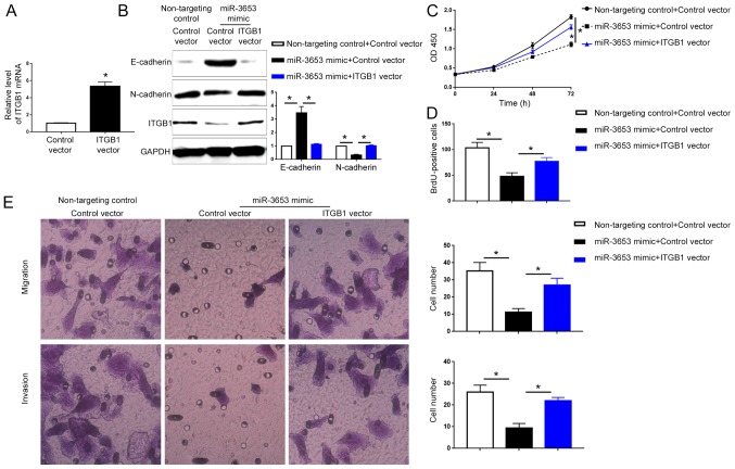
microRNAs (miRNAs) play critical roles in hepatocellular carcinoma (HCC). However, the expression and biological function of miR‑3653 in HCC remain unknown. The present study demonstrated that miR‑3653 expression was significantly decreased in HCC tissues and cells using qRT‑PCR. A decreased miR‑3653 level was associated with unfavorable clinical features and poor prognosis of HCC patients. MTT, BrdU, Transwell and western blot assays showed that miR‑3653 overexpression inhibited the growth, migration, invasion and epithelial‑mesenchymal transition (EMT) of HCCLM3 cells while its knockdown promoted the growth and metastatic ability of Hep3B cells. In vivo experiments showed that miR‑3653 overexpression inhibited the subcutaneous and the lung metastasis of HCCLM3 cells in nude mice. Mechanistically, integrin‑β1 (ITGB1) was identified to be the downstream target of miR‑3653 in HCC. ITGB1 overexpression reversed the inhibitory effects of miR‑3653 on the growth, metastasis and EMT of HCCLM3 cells.


Figures

Figure 1.
Figure 1.
Expression of miR-3653 in HCC tissues and cells. (A) The expression level of miR-3653 was compared between HCC tissues and adjacent non-tumor tissues. (B) The expression level of miR-3653 was compared between patients with large (>5 cm) and small (≤5 cm) tumors. (C) The expression level of miR-3653 was compared between patients with and without metastasis. (D) The expression of miR-3653 was evaluated in 4 HCC cell lines (Hep3B, Huh7, MHCC97H and HCCLM3) and immortalized hepatocyte L-02 cells. *P<0.05. HCC, hepatocellular carcinoma.
Figure 2.
Figure 2.
The prognostic value of miR-3653 in HCC patients. (A) Overall survival and (B) disease-free survival was compared between patients with a high miR-3653 level and those with a low miR-3653 level. *P<0.05. HCC, hepatocellular carcinoma.
Figure 3.
Figure 3.
miR-3653 overexpression inhibits the growth and metastatic ability of HCCLM3 cells. (A) Transfection of miR-3653 mimics significantly increased the expression level of miR-3653 in HCCLM3 cells. (B) miR-3653 overexpression decreased the cell viability of HCCLM3 cells, as suggested by MTT assay. (C) miR-3653 overexpression decreased the cell proliferation of HCCLM3 cells, as suggested by BrdU assay. (D) miR-3653 overexpression inhibited the migration and invasion of HCCLM3 cells, as suggested by Transwell assays. *P<0.05.
Figure 4.
Figure 4.
miR-3653 knockdown increases the growth and metastatic ability of Hep3B cells. (A) Transfection of miR-3653 inhibitor significantly decreased the expression level of miR-3653 in Hep3B cells. (B) miR-3653 knockdown increased the cell viability of Hep3B cells, as suggested by MTT assay. (C) miR-3653 knockdown increased the cell proliferation of Hep3B cells, as suggested by BrdU assay. (D) miR-3653 knockdown promoted the migration and invasion of Hep3B cells, as suggested by Transwell assays. *P<0.05.
Figure 5.
Figure 5.
miR-3653 inhibits the EMT in HCC cells. (A) Overexpression of miR-3653 led to an increased level of E-cadherin mRNA and decreased level of N-cadherin mRNA in HCCLM3 cells, as suggested by qRT-PCR. (B) miR-3653 overexpression led to an increased level of E-cadherin protein and decreased level of N-cadherin protein in HCCLM3 cells, as suggested by western blot analysis. (C) Knockdown of miR-3653 led to decreased level of E-cadherin mRNA and increased level of N-cadherin mRNA in Hep3B cells, as suggested by qRT-PCR. (D) miR-3653 knockdown led to decreased level of E-cadherin protein and increased level of N-cadherin protein in Hep3B cells, as suggested by western blot analysis. HCC, hepatocellular carcinoma. *P<0.05.
Figure 6.
Figure 6.
miR-3653 decreases the growth and metastasis of HCCLM3 cells in nude mice. (A) Overexpression of miR-3653 slowed down the growth of HCCLM3 cell-derived tumors in nude mice as suggested by subcutaneous injection experiment. Scale bar, 1 cm. (B) Overexpression of miR-3653 reduced the lung metastatic nodules formed by HCCLM3 cells in nude mice as suggested by tail vein injection experiment. Scale bar, 50 µm. *P<0.05.
Figure 7.
Figure 7.
ITGB1 is a direct target of miR-3653 in HCC. (A) miR-3653 was found to contain the complementary sequences mediating the binding between miR-3653 and ITGB1 3′-UTR. (B) Overexpression of miR-3653 decreased the luciferase activity of the wild-type (wt) ITGB1 3′-UTR but had no obvious influence on that of the mutated (mt) ITGB1 3′-UTR. Knockdown of miR-3653 significantly increased the luciferase activity of the wt 3′-UTR of ITGB1 but had no obvious influence on that of the mt 3′-UTR of ITGB1. (C) Overexpression of miR-3653 significantly decreased the mRNA level of ITGB1 in HCCLM3 cells. (D) Overexpression of miR-3653 significantly decreased the protein level of ITGB1 in HCCLM3 cells. (E) Knockdown of miR-3653 significantly increased the mRNA level of ITGB1 in Hep3B cells. (F) Knockdown of miR-3653 significantly increased the protein level of ITGB1 in Hep3B cells. (G) Immunohistochemical (IHC) staining for ITGB1 in HCC tissues with low miR-3653 levels and those with high miR-3653 levels. Scale bar, 100 µm. HCC, hepatocellular carcinoma; ITGB1; integrin-β1. *P<0.05.
Figure 8.
Figure 8.
ITGB1 mediates the biological functions of miR-3653 in HCC cells. (A) HCCLM3 cells were transfected with control vector or ITGB1 vector. qRT-PCR showed that the ITGB1 vector effectively increased ITGB1 mRNA in HCCLM3 cells. (B) HCCLM3 cells overexpressing miR-3653 were transfected with the control vector or ITGB1 vector. Transfection of ITGB1 vector restored ITGB1 expression in HCCLM3 cells overexpressing miR-3653, and led to decreased E-cadherin and increased N-cadherin. (C and D) Restoration of ITGB1 reversed the inhibitory effects of miR-3653 overexpression on the cell viability and proliferation of HCCLM3 cells. (E) Restoration of ITGB1 reversed the inhibitory effects of miR-3653 overexpression on the migration and invasion of HCCLM3 cells. Magnification, ×40. HCC, hepatocellular carcinoma; ITGB1; integrin-β1. *P<0.05.

References

    1. Yates LA, Norbury CJ, Gilbert RJ. The long and short of microRNA. Cell. 2013;153:516–519. doi: 10.1016/j.cell.2013.04.003. - DOI - PubMed
    1. Nelson KM, Weiss GJ. MicroRNAs and cancer: Past, present, and potential future. Mol Cancer Ther. 2008;7:3655–3660. doi: 10.1158/1535-7163.MCT-08-0586. - DOI - PubMed
    1. Lin K, Xu T, He BS, Pan YQ, Sun HL, Peng HX, Hu XX, Wang SK. MicroRNA expression profiles predict progression and clinical outcome in lung adenocarcinoma. Onco Targets Ther. 2016;9:5679–5692. doi: 10.2147/OTT.S111241. - DOI - PMC - PubMed
    1. Wu X, Li S, Xu X, Wu S, Chen R, Jiang Q, Li Y, Xu Y. The potential value of miR-1 and miR-374b as biomarkers for colorectal cancer. Int J Clin Exp Pathol. 2015;8:2840–2851. - PMC - PubMed
    1. El-Serag HB, Rudolph KL. Hepatocellular carcinoma: Epidemiology and molecular carcinogenesis. Gastroenterology. 2007;132:2557–2576. doi: 10.1053/j.gastro.2007.04.061. - DOI - PubMed

MeSH terms