Review
. 2019 Apr 19;431(9):1843-1868.
doi: 10.1016/j.jmb.2019.01.018. Epub 2019 Jan 18.

Metal Toxicity Links to Alzheimer's Disease and Neuroinflammation

Affiliations
Review

Metal Toxicity Links to Alzheimer's Disease and Neuroinflammation

Tee Jong Huat et al. J Mol Biol. .

Abstract

As the median age of the population increases, the number of individuals with Alzheimer's disease (AD) and the associated socio-economic burden are predicted to worsen. While aging and inherent genetic predisposition play major roles in the onset of AD, lifestyle, physical fitness, medical condition, and social environment have emerged as relevant disease modifiers. These environmental risk factors can play a key role in accelerating or decelerating disease onset and progression. Among known environmental risk factors, chronic exposure to various metals has become more common among the public as the aggressive pace of anthropogenic activities releases excess amount of metals into the environment. As a result, we are exposed not only to essential metals, such as iron, copper, zinc and manganese, but also to toxic metals including lead, aluminum, and cadmium, which perturb metal homeostasis at the cellular and organismal levels. Herein, we review how these metals affect brain physiology and immunity, as well as their roles in the accumulation of toxic AD proteinaceous species (i.e., β-amyloid and tau). We also discuss studies that validate the disruption of immune-related pathways as an important mechanism of toxicity by which metals can contribute to AD. Our goal is to increase the awareness of metals as players in the onset and progression of AD.

Keywords: dementia; environment; neurodegeneration; tau; β-amyloid.


Figures

Figure 1 ‒
Figure 1 ‒. Alzheimer’s disease causes and risks.
(Left panel) Rare mutations in app, psen1, and psen2 alter APP processing and are known strong genetic causes of AD. Other variationsin genes related to lipid metabolism, endocytosis and inflammatory responses, like apoe, trem2 and cd33, are more common in the population but they confer moderate to low risk to AD. The environmental risks for AD include aging, cardiac and metabolic disorders (i.e., diabetes and hypertension), level of education, reduced social engagement and severe traumatic brain injury. (Right panel) Neuropathologically, AD is characterized by the formation of Aβ plaques and NFTs in the brain. The production of Aβ occurs due to aberrant processing of the APP, whereby it is cleaved by β- and γ-secretases, instead of α-secretase. The Aβ peptide is prone to misfolding and aggregation, leading to eventual oligomerization and formation of Aβ plaques, which trigger a proinflammatory response from microglia and astrocytes. Aβ also causes the hyperphosphorylation of tau, leading to its dissociation from microtubules and their eventual destabilization within neurons. Hyperphosphorylated tau is also prone to aggregation, forming NFTs, which correlates with neuronal loss and neurodegeneration.
Figure 2 ‒
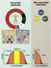
Figure 2 ‒. The molecular composition of the body: essential and non-essential elements.
In human biology, the constituents of the body are classified into four groups according to their increasing complexity: atomic, molecular, cellular and tissue-system. In this system, the more complex components are built by combining the more basic ones. At the atomic level, only four of the 118 chemical elements currently known (i.e., oxygen, carbon, hydrogen and nitrogen) are needed to make about 96% of the mass of the human body. Further 3.5% of body is composed of seven chemical elements, namely calcium, phosphorus, sulfur, potassium, sodium, chlorine and magnesium. The remaining constituents are trace elements. Of those, iron, zinc, manganese, copper, iodine, chromium, molybdenum, selenium and cobalt are considered essential nutrient elements for humans and are listed in order of recommended dietary allowance. Each of these elements has an optimal concentration in the body - too little or too much will result in reduced functionality or even death. In contrast, there are several non-essential elements likely to induce toxicity including aluminum, lead and cadmium. Non-essential elements can cause cellular dysfunction at low concentrations, followed by death if they persist in biological systems.
Figure 3 ‒
Figure 3 ‒. The impact of essential metals in AD.
(Left panel) In the healthy brain essential metals such as iron, copper, zinc and manganese are kept at a homeostatic level to ensure optimal cellular functions. To achieve these conditions, a complex mechanism exist to tightly regulate its intracellular and extracellular concentrations. For instance, iron (Fe2+) is kept within cells bound to the iron storage protein ferritin, making it available upon cellular needs. In neurons, APP can oxidize Fe2+ into Fe3+ inducing its release into the extracellular matrix. Once outside the cell, Fe3+ rapidly binds to the iron mobilization protein, transferrin, making iron accessible for further biological processes. APP is also involved in the conversion of Cu2+ into Cu+, favoring its removal from the brain. Zinc and manganese are also present in trace amounts which are finely regulated and essential to maintain brain function. (Right panel) In the AD brain, dyshomeostasis of essential metals seems to be linked with AD pathogenesis. Impairment of APP function, present in AD, can trigger an increased level of both intracellular and extracellular Fe2+ and Cu2+, and a reduction of extracellular Cu+, thus promoting its accumulation. Excessive Fe2+ and Cu2+ increases oxidative stress via production of ROS. In addition, iron, copper and zinc have higher binding affinity to Aβ and can promote its aggregation. Increased neuronal iron, copper and zinc also bind to tau protein and facilitate the formation of NFTs. Consequently, excessive amounts of heavy metals are found within plaques and NFT. Dyshomeostasis of essential metals in the extracellular space induce microglial and astrocytic activation, followed by the overproduction of proinflammatory cytokines such as, IL-1β and TNF-α.
Figure 4 ‒
Figure 4 ‒. The impact of non-essential metals in AD.
The presence of non-essential metals such as lead, aluminum, and cadmium have neurotoxic effects on the brain which are exacerbated in AD. Lead exposure increases APP and BACE1 expression and disrupts microglial functioning, together increasing Aβ production and plaque formation. Increased intracellular aluminum competes for the iron binding site in the iron-responsive element; as a result, Fe2+ accumulates and increases the production of ROS. Aluminum also accumulates in NFT-bearing neurons. In addition, aluminum can bind to Aβ and induce its aggregation. Cadmium also binds to Aβ and involved in the formation of plaques. Heavy metal exposure induces microglial and astrocytic activation and subsequent increase in production of proinflammatory proteins, including IL-1β, IL-8, TNF-α, IL-6 and iNOS. Extracellular levels: changes in metal levels in the extracellular space include the metals accumulated in plaques, note that it might not necessarily reflect the number of free ions available in the extracellular space.

References

    1. United Nations, Department of Economic and Social Affairs, Population Division (2017). World Population Prospects: The 2017 Revision, Key Findings and Advance Tables.
    1. Livingston G, Sommerlad A, Orgeta V, Costafreda SG, Huntley J, Ames D, Ballard C, Banerjee S, Burns A, Cohen-Mansfield J, Cooper C, Fox N, Gitlin LN, Howard R, Kales HC, Larson EB, Ritchie K, Rockwood K, Sampson EL, Samus Q, Schneider LS, Selbaek G, Teri L & Mukadam N (2017). Dementia prevention, intervention, and care. Lancet. 390,2673–734. - PubMed
    1. Newcombe EA, Camats-Perna J, Silva ML, Valmas N, Huat TJ & Medeiros R (2018). Inflammation: The link between comorbidities, genetics, and alzheimer’s disease. J Neuroinflammation. 15,276. - PMC - PubMed
    1. Plummer S, Van Den Heuvel C, Thornton E, Corrigan F & Cappai R (2016). The neuroprotective properties of the amyloid precursor protein following traumatic brain injury. Aging Dis. 7,163–79. - PMC - PubMed
    1. Bateman RJ, Xiong C, Benzinger TL, Fagan AM, Goate A, Fox NC, Marcus DS, Cairns NJ, Xie X, Blazey TM, Holtzman DM, Santacruz A, Buckles V, Oliver A, Moulder K, Aisen PS, Ghetti B, Klunk WE, Mcdade E, Martins RN, Masters CL, Mayeux R, Ringman JM, Rossor MN, Schofield PR, Sperling RA, Salloway S & Morris JC (2012). Clinical and biomarker changes in dominantly inherited alzheimer’s disease. N Engl J Med. 367,795–804. - PMC - PubMed

Publication types

MeSH terms