

Case Reports
doi: 10.1016/j.jaci.2018.12.1012.
Epub 2019 Jan 18.
Novel proteasome assembly chaperone mutations in PSMG2/PAC2 cause the autoinflammatory interferonopathy CANDLE/PRAAS4
Affiliations
- PMID: 30664889
- PMCID: PMC6565382
- DOI: 10.1016/j.jaci.2018.12.1012
Item in Clipboard
Case Reports
Novel proteasome assembly chaperone mutations in PSMG2/PAC2 cause the autoinflammatory interferonopathy CANDLE/PRAAS4
J Allergy Clin Immunol.
2019 May.
Abstract
Chronic Atypical Neutrophilic Dermatosis with Lipodystrophy and Elevated temperature (CANDLE) is a rare autoinflammatory interferonopathy caused by additive loss-of-function mutations in proteasome genes. Mutations in the proteasome chaperone, PSMG2/PAC2 are a novel cause of CANDLE.
Conflict of interest statement
Conflicts of Interest
The authors declare no relevant conflict of interest. Dr. Goldbach-Mansky has received grant support from SOBI, Regeneron, Novartis and Eli Lilly.
Figures
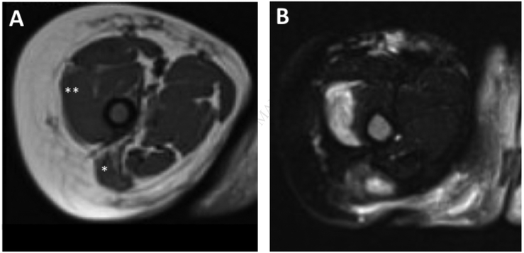
(A) T1 weighted axial MRI of right and left thigh shows atrophy and fatty replacement of the gluteus maximus and vastus medialis muscles. (B) The corresponding T2 weighted fat suppressed axial image shows patchy enhancement in these areas suggestive of active inflammation.

(A) Visualization of the binary alignment map (bam) using Integrative Genomics Viewer (IGV) revealed that the two variants are in trans configuration. The sequencing reads containing the 2 base-pair deletion lack the missense mutation (red dashed box), whereas the sequencing reads containing the missense mutation lack the 2 base-pair deletion (blue dashed box), thus suggesting that each variant was inherited from a different parent which was confirmed by Sanger sequencing. (B) Multispecies alignment of the PSMG2 protein region affected by the compound heterozygous variants confirms conservation across species for both variants.
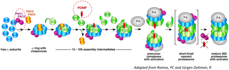
The PAC1/PAC2 heterodimer initiates α-subunit-ring assembly by binding to the20S core protein subunits, α5/PSMA5 and α4/PSMA7 and prevents dimerization of the α-rings and promotes the assembly of the hetero-heptameric αβ-subunit-ring (Le Tallec et al 2007). The PAC3/PAC4 complex binds later and dissociates from the maturing proteasome complex, at the “half-mer (−β7) intermediate” stage when only the β7-subunit is lacking. At that point, POMP is recruited to mediate incorporation of either standard β5/PSMB5 or the β5i/LMP7/PSMB8 immunoproteasome subunit and dimerization of two “half-proteasome precursor complexes” to form the short-lived “nascent proteasome). This conformational change triggers the release of the PAC1/PAC2 complex from the nascent proteasome and enables binding of the 19S regulatory particle.,, In the red circles: Loss of function mutations in PSMG2/PAC2 (compound heterozygous, disease-causing mutations lead to additive loss of function) and POMP/POMP ((alias UMP1), heterozygous, disease-causing mutations cause haploinsufficiency), mutations in both genes cause CANDLE/PRAAS.
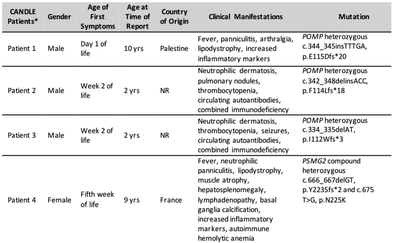
*denotes CANDLE patients with currently known chaperone mutations in POMP and including our patient with PSMG2 mutations. NR: not reported (in this figure, cases from refs. and were combined)
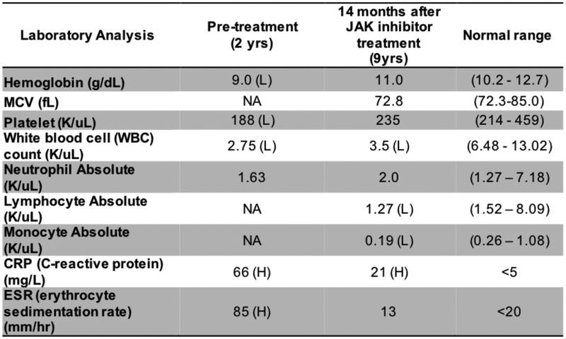
H: high; L: low; NA: not available
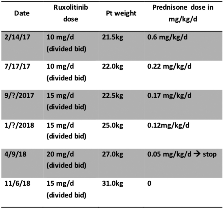
Ruxolitinib dose, weight changes and steroid taper on JAK inhibitor baricitinib.
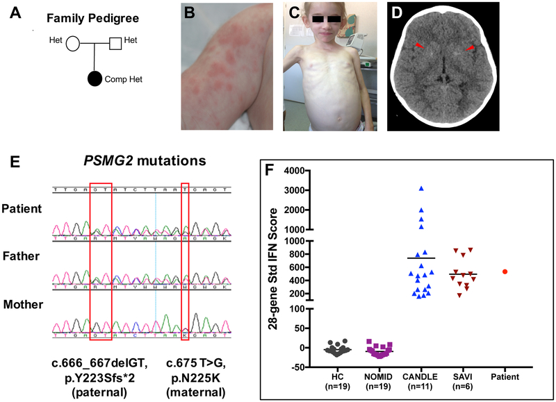
A, Family tree depicting that both patient’s parents are heterozygous for a different PSMG2 mutation. B, Nodular skin rash at the age of 7months. C, Generalized lipoatrophy involving the face and upper extremities at the age of 7 years. D, A brain CT showing faint bilateral basal ganglia calcifications (arrow heads). E, Sanger sequencing electropherogram from patient and parents showing that the frameshift deletion is on the paternal allele and the missense variant is on the maternal allele. F, 28-gene interferon (IFN) score performed with a customized Nanostring assay demonstrated that our patient has a high IFN score, similar to the observed in patients with the interferonopathies CANDLE and SAVI. Mean ± SD of 28-gene IFN score: healthy controls (HC): −4.8 ± 9.53; neonatal onset multisystem inflammatory disease (NOMID): −9.37 ± 10.97; CANDLE: 738.2 ± 754.4; SAVI: 495.6 ± 225.9; Patient: 533.6

A, Fibroblasts lysates from the patient (Pt.) and CANDLE/PRAAS disease controls (DC) had approximately 75% of proteasome activity compared to healthy controls (HCs). Means±SD estimated from triplicates and normalized against HC1+2 (n=3). B, PRAAS patients accumulated insoluble ubiquitin aggregates. C, Reduced PAC1 and PAC2 expression in patient’s sample compared to HCs and DCs. Expression of subunits POMP/POMP, α6/PSMA1, β5i/PSMB8, PA28b/PSME2, RPT6/PSMC5, PA200/PSME4 was normal. β–Tubulin: loading control. D, Native PAGE-immunoblot-analyses shows a reduced PAC1 and PAC2 incorporation into the proteasome complexes. Reduced α6 staining indicates less proteasome complexes in all CANDLE/PRAAS patients E, Overlay of chymotrypsin-like activity shows reduced proteolytic activity in the main proteasome complexes for CANDLE/PRAAS patients.. F, The epitope–tagged PAC2 variants (wildtype (WT); fs (frameshift mutation); NK (N225K mutation) were expressed in HeLa cells and detected by a Flag-specific antibody. MOCK, empty vector backbone; HeLa, non-transfected control. β–Tubulin: loading control. Neomycin-Phosphatase (Neo-P): transfection efficiency control. All PAC2-variant mRNAs were equally expressed in HeLa cells (RT-PCR). GAPDH: loading control. Panels A-F: representative results from n=3. G, Density gradient fractionation (panel F) shows incorporation of both mutant Pac2 variants into proteasome complexes, without impacting total proteasome formation (α6 staining).
References
-
- Liu Y, Ramot Y, Torrelo A, Paller AS, Si N, Babay S, et al. Mutations in proteasome subunit beta type 8 cause chronic atypical neutrophilic dermatosis with lipodystrophy and elevated temperature with evidence of genetic and phenotypic heterogeneity. Arthritis Rheum 2012;64(3):895–907. - PMC - PubMed
-
- Ciechanover A. The ubiquitin-proteasome proteolytic pathway. Cell 1994;79(1):13–21. - PubMed
-
- Seifert U, Bialy LP, Ebstein F, Bech-Otschir D, Voigt A, Schroter F, et al. Immunoproteasomes preserve protein homeostasis upon interferon-induced oxidative stress. Cell 2010;142(4):613–24. - PubMed
Publication types
MeSH terms
Substances
Supplementary concepts
Grants and funding
LinkOut - more resources
Full Text Sources
Molecular Biology Databases
