. 2019 Jan 2;60(1):322-330.
doi: 10.1167/iovs.18-25667.

Selective Ablation of Megalin in the Retinal Pigment Epithelium Results in Megaophthalmos, Macromelanosome Formation and Severe Retina Degeneration

Affiliations

Selective Ablation of Megalin in the Retinal Pigment Epithelium Results in Megaophthalmos, Macromelanosome Formation and Severe Retina Degeneration

Tina Storm et al. Invest Ophthalmol Vis Sci. .

Abstract

Purpose: Mutations in the megalin-encoding gene, LRP2, cause high myopia as seen in patients suffering from Donnai-Barrow/facio-oculo-acoustico-renal syndrome. Megalin is present in both the nonpigmented epithelium of the ciliary body and in the RPE. In this study, we set out to establish an animal model to study the mechanisms underlying the ocular phenotype and to establish if high myopia/megaophthalmos is induced by postnatal megalin-deficiency in the RPE.

Methods: Postnatal RPE-specific deletion of megalin was generated by crossing mice bearing a homozygous loxP-flanked Lrp2 allele with transgenic mice expressing the Cre recombinase driven by the BEST1 promotor. The model was investigated by immunohistologic techniques, and transmission electron microscopy.

Results: Mice with postnatal RPE-specific loss of megalin developed a megaophthalmos phenotype with dramatic increase in ocular size and severe retinal thinning associated with compromised vision. This phenotype was present at postnatal day 14, indicating rapid development in the period from onset of BEST1 promotor activity at postnatal day 10. Additionally, RPE melanosomes exhibited abnormal size and morphology, suggested by electron tomography to be caused by fusion events between multiple melanosomes.

Conclusions: Postnatal loss of megalin in the RPE induces dramatic and rapid ocular growth and retinal degeneration compatible with the high myopia observed in Donnai-Barrow patients. The morphologic changes of RPE melanosomes, believed to be largely inert and fully differentiated at birth, suggested a continued plasticity of mature melanosomes and a requirement for megalin to maintain their number and morphology.


Figures
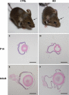
Figure 1
Figure 1
Overall eye size in control and mutant mice. Control (A) and mutant (B) eyes shown in vivo before enucleation and preparation. Mutant eyes are clearly protruding and enlarged compared to control eyes. Representative histologic cross-sections of enucleated eyes from 14-day-old (C, D) and adult (14–17.5 months old; E, F) normal (C, E) and mutant eyes (D, F) shows a large difference in the overall eye size between the two groups. Scale bars: C, D, E, and F = 1 mm.
Figure 2
Figure 2
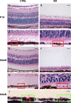
Retinal morphology in 14-day-old and adult normal and mutant eyes. Hematoxylin-eosin–stained histologic cross-section of control (CTRL, left panel) and mutant (KO, right panel) mouse eyes shows severe thinning of all retinal layers in both 14-day-old (B) and adult (14–17.5 months old) (F) mutant mice. Enlargements of the RPE in these mice are given directly below (C, D, G, H) and shows that RPE melanosomes of mutant retinas are abnormally large and reduced in number (melanosomes highlighted by red squares). (I) Immunohistochemical detection of the Cre recombinase in mutant retinas showed a mosaic expression pattern (Cre recombinase positive nuclei are red stained, highlighted by green squares) and also that expression of the Cre recombinase correlates with the presence of macromelanosomes. Scale bars: A, B, E, F = 25 μm and C, D, G, H, I = 5 μm.
Figure 3
Figure 3
Immunohistochemical localization of megalin in control and mutant eyes. (A–D) Whole eye sections labeled with anti-megalin (green) and propidium iodide (red). Megalin is detected throughout the retina pigment epithelium in control mouse eyes (A) but only in a mosaic pattern in mutant eyes (B). Strong megalin expression is seen in the CMZ of both control (C) and mutant (D) eyes. RPE nuclei are indicated with white asterisks. ONL, outer nuclear layer; CMZ, ciliary marginal zone. Scale bars: A, B, C, and D = 10 μm.
Figure 4
Figure 4
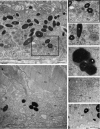
RPE ultrastructure in control and mutant mice. Transmission electron micrographs of the outermost retinal layers in control (CTRL, left panel) and mutant mice (KO, right panel) shows no apparent ultrastructural differences in 5-day-old mice (A, B). In contrast, we observed a large number of ultrastructural abnormalities in 14-day-old mutant mice (D) including thinning of the photoreceptor outer segment layer, macromelanosomes, melanolipofuscin-like structures, as well as a loss of basal infoldings. Scale bars: A, B, C, and D = 2 μm.
Figure 5
Figure 5
RPE melanosome ultrastructure in 14-day-old mutant mice. High magnification transmission electron micrographs showing the abnormal size and multispherical shapes of selected macromelanosomes from 14-day-old mutant RPE cells (A, B; n = 4). Still frame from a 3D tomographic analysis (see Supplementary Video 1) of a thick epon section from a 14-day-old mutant RPE cell (C). The multispherical shape of the macromelanosome is surrounded by a single limiting membrane encompassing the entire structure (highlighted in green) and is comprised of three separate melanosomes, each identifiable by the perimeters of individual packed melanin cores (highlighted in red [D]). Scale bars: A, B = 1 μm and C, D = 200 nm.
Figure 6
Figure 6
RPE cell ultrastructure in control and mutant 14-day-old mice. Transmission electron micrographs of RPE cells from control (A, C, [zoom of region in A] and D) and mutant (B, E, F, G) 14-day-old mice illustrating the ultrastructural abnormalities identified in mutant mice including loss of basal infoldings (B), large melanolipofuscin-like structures (B, E), distended and fragmented endoplasmic reticulum throughout the cytoplasm (B, F, G), scattered and detached ribosomes (example indicated by small black box [F, G]), apparent increased number of Golgi apparatus (G) as well as a general absence of clathrin-coated vesicles (B, F, G). BI, basal infoldings; BM, basal membrane; ccv, clathrin-coated vesicles; dER, dilated endoplasmic reticulum; G, Golgi apparatus; M, melanosome; Mit, mitochondrion; ML, melanolipofuscin-like; POS, photoreceptor outer segment; Pr, polyribosome. Scale bars: A, B, G = 1 μm, C, D = 500 nm; E, F = 200 nm.

Similar articles

Cited by

References

    1. Pober BR, Longoni M, Noonan KM. A review of Donnai-Barrow and facio-oculo-acoustico-renal (DB/FOAR) syndrome: clinical features and differential diagnosis. Birth Defects Res A Clin Mol Teratol. 2009;85:76–81. - PMC - PubMed
    1. Kantarci S, Al-Gazali L, Hill RS, et al. Mutations in LRP2, which encodes the multiligand receptor megalin, cause Donnai-Barrow and facio-oculo-acoustico-renal syndromes. Nat Genet. 2007;39:957–959. - PMC - PubMed
    1. Patel N, Hejkal T, Katz A, Margalit E. Ocular manifestations of Donnai-Barrow syndrome. J Child Neurol. 2007;22:462–464. - PubMed
    1. Kantarci S, Ragge NK, Thomas NS, et al. Donnai-Barrow syndrome (DBS/FOAR) in a child with a homozygous LRP2 mutation due to complete chromosome 2 paternal isodisomy. Am J Med Genet A. 2008;146A:1842–1847. - PMC - PubMed
    1. Schrauwen I, Sommen M, Claes C, et al. Broadening the phenotype of LRP2 mutations: a new mutation in LRP2 causes a predominantly ocular phenotype suggestive of Stickler syndrome. Clin Genet. 2013;86:282–286. - PubMed

Publication types