. 2019 Jan 21;19(1):94.
doi: 10.1186/s12885-019-5299-8.

Glucocorticoids promote the development of azoxymethane and dextran sulfate sodium-induced colorectal carcinoma in mice

Affiliations

Glucocorticoids promote the development of azoxymethane and dextran sulfate sodium-induced colorectal carcinoma in mice

Bo Li et al. BMC Cancer. .

Abstract

Background: Stress has been suggested as a promoter of tumor growth and development. Glucocorticoids (GCs) are the main stress hormones and widely prescribed as drugs. However, the effect of GCs on the development and progression of colorectal carcinoma (CRC) is unclear.

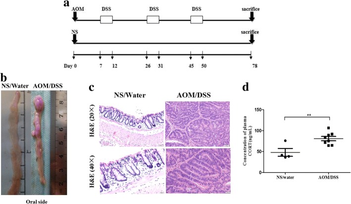
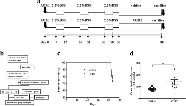
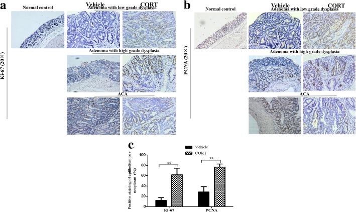
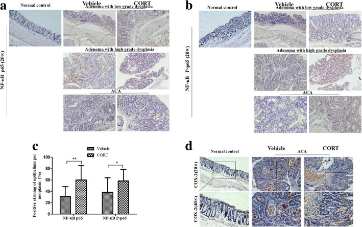
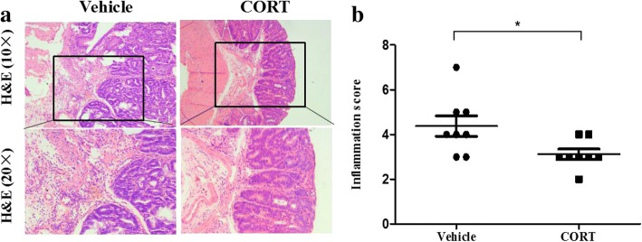
Methods: We evaluated the effect of corticosterone (CORT) on azoxymethane and dextran sulfate sodium (AOM/DSS)-induced carcinogenesis in the colorectum of C57BL/6 strain mice. Plasma level of CORT was detected by radioimmunoassay. The expression of proliferation markers (Ki-67 and PCNA), nuclear factor (NF)-κB p65 and phosphoto-p65 (P-p65), as well as cyclooxygenase (COX)-2 were determined by immunohistochemistry. Inflammation in colorectum was evaluated by histopathology.

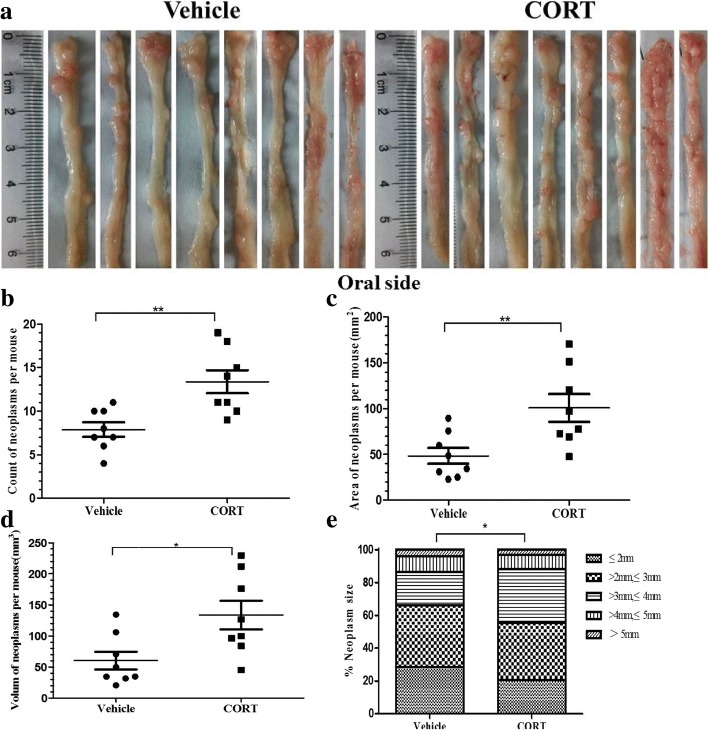
Results: CORT feeding in drinking water of mice not only significantly elevated plasma CORT concentration, but also significantly increased the incidence and neoplasms burden (number and size of neoplasms) in colorectum. CORT also significant enhanced the expression of cell proliferation marker (Ki-67 and PCNA), NF-κB p65 and P-p65 as well as COX-2 in colorectal neoplasm of AOM/DSS-treated mice.

Conclusion: In this study, we have found for the first time that CORT at stress level potentially promotes the growth and development of AOM/DSS-induced colorectal adenoma and carcinoma in mice. Up-regulation of NF-κB and COX-2 may be involved in the promoting effect of CORT.

Keywords: Azoxymethane/dextran sodium sulfate; Colorectal carcinoma; Corticosterone; Nuclear factor-κB; Tumor development.


Conflict of interest statement

Ethics approval

All animal experiments were approved by the Animal Care and Use Committee of Second Military Medical University (Shanghai, China) and performed in compliance with the University’s Guidelines for the Care and Use of Laboratory Animals (reference number: 20131015001).

Consent for publication

Not applicable.

Competing interests

The authors declare that they have no competing interests.

Publisher’s Note

Springer Nature remains neutral with regard to jurisdictional claims in published maps and institutional affiliations.

Figures

Fig. 1
Fig. 1
AOM/DSS treatment induces CRC in C57BL/6 male mice successfully and increases the plasma CORT of model mice. The scheme shows one time administration of AOM followed by three cycles of DSS treament and separated by 14-day recovery periods. Colon was examined 28 days after the third cycle of DSS treatment (a). Representative longitudinal views of colorectum from control (NS/Water) mice and model (AOM/DSS) mice (b). Pathological examination confirmed the most neoplasms of model mice as adenocarcinoma (c). Plasma CORT of model mice (number = 8) and control mice (number = 4) were tested by radioimmunoassay (d). Data expressed as mean ± SD. **P < 0.01 by unpaired t-test analysis. AOM azoxymethane, DSS dextran sodium sulphate, CRC colorectal carcinoma, NS normal saline, CORT corticosterone
Fig. 2
Fig. 2
CORT feeding increases plasma CORT of AOM/DSS-induced CRC mice. The scheme shows model mice recovered for 7 days after the last cycle of DSS treatment and then undertook with vehicle treatment (VT, 0.5% ethanol, number = 10) or CORT treatment (CT,50 mg/L, number = 11) randomly for 31 days (a). Methodology workflow of the study (b). Survival rate of CT mice (number = 11) and VT mice (number = 10) (c). Plasma CORT of CT mice (number = 8) compared with VT mice (number = 8) (d). Data expressed as mean ± SD. *P < 0.05, **P < 0.01 by unpaired t-test analysis. CORT corticosterone, CT CORT treatment, VT vehicle treatment
Fig. 3
Fig. 3
CORT feeding increases development of AOM/DSS-induced CRC in mice. Representative longitudinal views of colorectum from the VT mice and the CT mice (a). Count of neoplasms per mouse (b), area of neoplasms per mouse (c) and volume of neoplasms per mouse (d) of the VT and the CT mice. Percentage of neoplasms per size group for each treatment mice (e). Number = 8 per group. Data expressed as mean ± SD. *P < 0.05, **P < 0.01 by unpaired t-test and two-way ANOVA for neoplasms size distribution analysis. Length and width of neoplasms were measured by electronic vernier caliper. Area = [(length + width)*0.5]2, Volume = length*width2*0.52. CT CORT treatment, VT vehicle treatment
Fig. 4
Fig. 4
CORT feeding elevates expression of proliferation marker in AOM/DSS-induced CRC mice. The colorectum of model mice with VT or CT (number = 3 per group) were processed for immunostaining of proliferation marker. Representative images of normal colorectal epithelia (Normal control) and neoplasms area, classified pathologically as adenoma with low grade dysplasia, adenoma with high grade dysplasia and ACA, stained with antibody of Ki-67 (a, at 20X), PCNA (b, at 20X) and their bar diagrams showing the percentages of Ki-67 and PCNA positive colonic epithelial cells (c). Data expressed as mean ± SD. **P<0.01 by unpaired t-test analysis. CT CORT treatment, VT vehicle treatment
Fig. 5
Fig. 5
CORT feeding elevates expression of NF-κB and COX-2 in AOM/DSS-induced CRC in mouse. The colons of model mice with CT or VT (number = 6 per group) were processed for immunostaining of NF-κB and COX-2. Representative images of normal colorectal epithelia (Normal control) and neoplasms area, classified pathologically as adenoma with low grade dysplasia, adenoma with high grade dysplasia and ACA, stained with antibody of NF-κB p65 (a, at 20X), NF-κB P-p65 (b, at 20X) and their bar diagrams showing the percentages of NF-κB p65 and P-p65 (c) positive cells of different treatment groups. Representative images of normal colorectal epithelia (Normal control) and ACA area stained with antibody of COX-2 (d, at 20X and 40X). Data expressed as mean ± SD.*P<0.05, **P<0.01 by t-test for IHC data. ACA adenocarcinoma, IHC immunohistochemistry, CT CORT treatment, VT vehicle treatment
Fig. 6
Fig. 6
CORT feeding inhibits inflammation of neoplasms from AOM/DSS-induced CRC mice. The colorectum of model mice with CT or VT (number = 8 per group) were processed for H&E staining. Representative images of neoplasm area (a, at 10X and 20X). Semi-quantitative histological measurement of total inflammation (inflammation score) were calculated by adding the values for percent ulceration, percent re-epithelialization, and active, chronic and transmural inflammation (b). Data expressed as mean ± SD. *P<0.05 by unpaired t-test analysis. H&E hematoxylin and eosin, CORT corticosterone, CT CORT treatment, VT vehicle treatment

References

    1. Neeman E, Ben-Eliyahu S. Surgery and stress promote cancer metastasis: new outlooks on perioperative mediating mechanisms and immune involvement. Brain Behav Immun. 2013;30(Suppl):32–40. doi: 10.1016/j.bbi.2012.03.006. - DOI - PMC - PubMed
    1. Moreno-Smith M, Lutgendorf SK, Sood AK. Impact of stress on cancer metastasis. Future Oncol. 2010;6(12):1863–1881. doi: 10.2217/fon.10.142. - DOI - PMC - PubMed
    1. Gundisch S, Boeckeler E, Behrends U, Amtmann E, Ehrhardt H, Jeremias I. Glucocorticoids augment survival and proliferation of tumor cells. Anticancer Res. 2012;32(10):4251–4261. - PubMed
    1. Volden PA, Conzen SD. The influence of glucocorticoid signaling on tumor progression. Brain Behav Immun. 2013;30(Suppl):26–31. doi: 10.1016/j.bbi.2012.10.022. - DOI - PMC - PubMed
    1. Reiche EM, Nunes SO, Morimoto HK. Stress, depression, the immune system, and cancer. Lancet Oncol. 2004;5(10):617–625. doi: 10.1016/S1470-2045(04)01597-9. - DOI - PubMed