

Agreement study between color and IR retinal images based on retinal vasculature morphological parameters
- PMID: 30665394
- PMCID: PMC6341663
- DOI: 10.1186/s12886-018-0997-6
Agreement study between color and IR retinal images based on retinal vasculature morphological parameters
Abstract
Background: Color fundus photography have been extensively used to explore the link between retinal morphology changes associated with various disease i.e. Diabetic Retinopathy, Glaucoma. The development of multimodal imaging system that integrates Infrared Scanning Laser Ophthalmoscope (IR-SLO) and Optical Coherence Tomography (OCT) could help in studying these diseases at an early stage. The aim of this study was to test the agreement between the retinal vasculature parameters from the Infrared images obtained from optical coherence tomography and color fundus imaging.
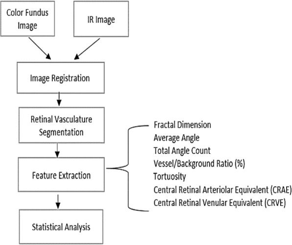
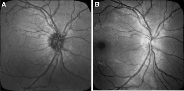
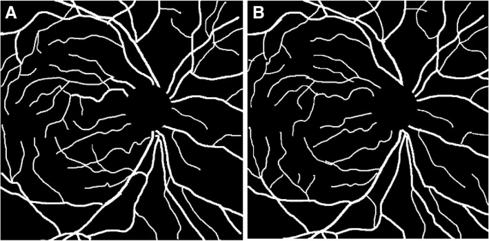
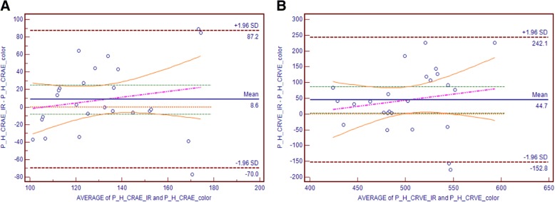
Methods: The IR and Color retinal images were obtained from 16 volunteer participants and seven retinal vessel parameters, i.e. Fractal Dimension (FD), Average Angle (ABA), Total Angle Count (TAC), Tortuosity (ST), Vessel/Background ratio (VBR), Central Retinal Arteriolar Equivalent (CRAE) and Central Retinal Venular Equivalent (CRVE) were extracted from these retinal images using Retinal Image Vasculature Assessment software (RIVAS) and Integrative Vessel Analysis (IVAN).
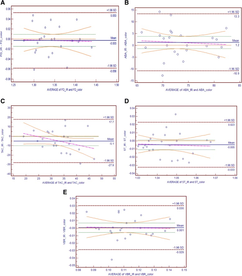
Results: The Bland Altman plot was used to investigate the agreement between the two modalities. The paired sample t-test was used to assess the presence of fixed bias and the slope of Least Square Regression (LSR) line for the presence of proportional bias. The paired sample t-test showed that there was no statistically significant difference between Color and IR based on retinal vessel features (all p values > 0.05). LSR also revealed no statistically significant difference in the retinal vessel features between Color and IR.
Conclusion: This study has revealed that there is a fair agreement between Color and IR images based on retinal vessel features. This research has shown that it is possible to use IR images of the retina to measure the retinal vasculature parameters which has the advantage of being flash-less, can be used even if there is opacity due to cataract, and can be performed along with OCT on the same device.
Keywords: Infrared scanning laser ophthalmoscope; Integrative vessel analysis; Optical coherence tomography; Retinal image vasculature assessment software.
Conflict of interest statement
Ethics approval
The experimental protocol was approved by RMIT University human experiments ethics sub-committee and performed in accordance with Helsinki accord 1986 (modified 2004). The experimental protocol was described in plain language to each participant and written consent was obtained prior to the experiment.
Competing interests
The authors declare that they have no competing interests.
Publisher’s Note
Springer Nature remains neutral with regard to jurisdictional claims in published maps and institutional affiliations.
Figures
References
-
- Kumar D, Aliahmad B, Arjunan SP. Fractals: applications in biological Signalling and image processing: Taylor & Francis; 2016.
-
- Jain AB, Prakash VJ, Bhende M. Techniques of fundus imaging, vol. 33. India: Scientific Journal of Medical & Vision Research Foundations; 2015.
MeSH terms
LinkOut - more resources
Full Text Sources
