Review
. 2019 Apr;27(4):303-322.
doi: 10.1016/j.tim.2018.12.009. Epub 2019 Jan 19.

Fighting Staphylococcus aureus Biofilms with Monoclonal Antibodies

Affiliations
Review

Fighting Staphylococcus aureus Biofilms with Monoclonal Antibodies

Dina Raafat et al. Trends Microbiol. 2019 Apr.

Abstract

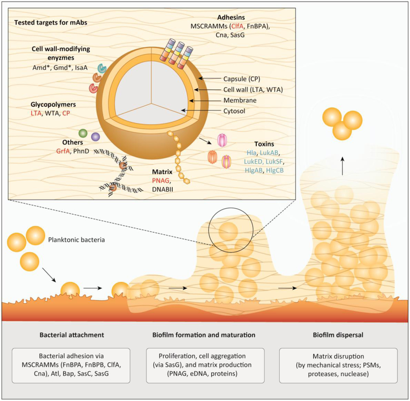
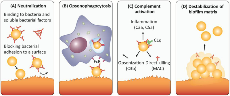
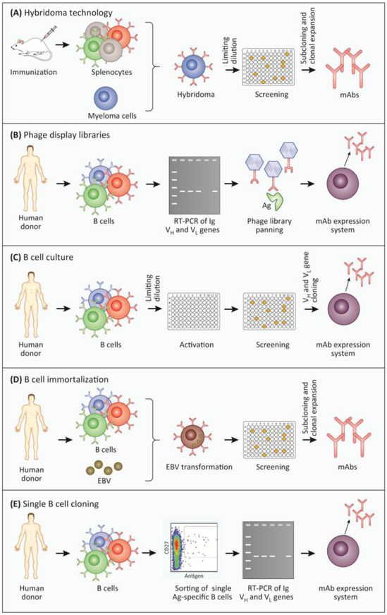
Staphylococcus aureus (S. aureus) is a notorious pathogen and one of the most frequent causes of biofilm-related infections. The treatment of S. aureus biofilms is hampered by the ability of the biofilm structure to shield bacteria from antibiotics as well as the host's immune system. Therefore, new preventive and/or therapeutic interventions, including the use of antibody-based approaches, are urgently required. In this review, we describe the mechanisms by which anti-S. aureus antibodies can help in combating biofilms, including an up-to-date overview of monoclonal antibodies currently in clinical trials. Moreover, we highlight ongoing efforts in passive vaccination against S. aureus biofilm infections, with special emphasis on promising targets, and finally indicate the direction into which future research could be heading.

Keywords: Staphylococcus aureus; biofilm; infection; monoclonal antibodies; passive vaccination; vaccine.


Figures

Figure 1:
Figure 1:. Overview on tested targets for an antibody-based preventive or therapeutic strategy against biofilm-associated S. aureus infections.
Main figure: Biofilm formation in staphylococci comprising three main stages: bacterial attachment to a surface, biofilm formation and maturation, and biofilm detachment / dispersal. For the attachment to (a)biotic surfaces, S. aureus relies on a broad spectrum of functionally redundant adhesins such as the MSCRAMMs (ClfA, Cna, FnbA, FnbB). After successful adhesion, bacteria start proliferation and production of the biofilm matrix consisting of eDNA, stabilized by DNABII, PNAG and proteins. Eventually, biofilm dispersal is mediated by mechanical shear stress (e.g. in a blood vessel) or by dispersion factors like PSMs, nuclease, and proteases. Insert: Molecular targets for antibody based therapies tested in preclinical in clinical studies include adhesins and cell-wall modifying enzymes and other cell wall-attached proteins, surface glycopolymers, biofilm matrix components, as well as toxins and immune evasion proteins. Targets from preclinical studies, ongoing clinical trials and failed clinical trials are shown in black, blue and red, respectively. The asterisk indicates that the S. aureus protein autolysin (Atl) is proteolytically processed into two enzymes, autolysin amidase (Amd) and autolysin glucosaminidase (Gmd), which stay non-covalently attached to the cell surface. Abbreviations: Atl (autolysin); Amd (autolysin amidase); Bap (biofilm-associated protein ); ClfA (clumping factor A); Cna (collagen-binding protein); CP (capsular polysaccharides); DNABII (DNABII family proteins); eDNA (extracellular DNA); FnBPA/FnBPB (fibronectin-binding protein A and B); Gmd (autolysin glucosaminidase); GrfA (ABC transporter); Hla (α-toxin); Hlg (γ-haemolysin); IsaA (Immunodominant staphylococcal antigen A); LTA (lipoteichoic acid); Luk (Leukotoxins); mAb (monoclonal antibody); MSCRAMM (microbial surface components recognizing adhesive matrix molecule); PhnD (subunit of alkylphosphonate ABC transporter); PNAG (poly-N-acetyl-ß-(1,6)-glucosamine); PSMs (phenol soluble modulins); Sasc/G (S. aureus surface protein C and G); WTA (wall teichoic acid).
Figure 2:
Figure 2:. Antibodies can interfere with biofilm formation and promote dispersal of established biofilms by several mechanisms.
A) Secreted staphlyococcoal proteins (e.g. immune evasion molecules, toxins, exoenzymes) as well as surface proteins are involved in biofilm development and are hence potential targets for therapeutic purposes. High affinity IgA and IgG antibodies can neutralize the action of bacterial toxins and surface proteins. Moreover, antibodies can bind to bacterial adhesins (e.g. ClfA, FnBPA) and cell wall components (e.g. PNAG), thereby blocking initial attachment to host matrices and subsequent initiation of biofilm formation. B) Surface-bound antibodies (most prominently IgG) can trigger the uptake and destruction by neutrophils and macrophages expressing Fc receptors (FcR) on their surface (opsonophagocytosis). Activation of neutrophils can also trigger granule release, oxidative burst and NETosis. C) Surface-bound antibodies (IgM and IgG) trigger complement activation via the classical pathway. Following binding of C1q to the surface-bound antibody, the complement cascade is initiated resulting in the formation of the C3 convertase, which cleaves the central component of all complement pathways, C3, into C3a and C3b. C3b acts as an opsonin, enabling phagocytes that express the C3b receptor to ingest C3b-coated bacteria more easily. The soluble C3a (as well as C5a) act as chemo-attractants that recruit immune cells to the site of infection causing inflammation. C3 activation also triggers the formation of the membrane attack complex (MAC) that generates lytic pores in certain pathogens. Gram-positive bacteria, including S. aureus, are protected from MAC-dependent lysis by their thick peptidoglycan layer [132]. D) Antibodies targeting different components of the biofilm matrix, e.g. DNABII, can destabilize a biofilm matrix and thereby promote bacterial dispersal and clearance by immune cells or antibiotics. Abbreviations: FcR (Fc receptor); MAC (membrane attack complex).
Figure 3:
Figure 3:. Methods commonly used for the generation of monoclonal antibodies (mAbs).
A) Hybridoma technology. Following immunization with an antigen, mice start producing large amounts of antigen-specific B cells. These cells are harvested from the spleen and fused with myeloma cells. The resulting hybridoma cells are screened for the secretion of antigen-specific antibodies. Antigen-specific hybridoma cells are selected by limiting dilution (subcloning) [3]. B) Phage display library. Initially, mRNA is isolated from B cells or plasma cells and then reverse-transcribed into cDNA. The variable light and heavy chains are amplified via PCR and ligated into a phage display vector. The resulting phage library consists of 108-1010 different phages, each encoding a single surface-expressed mAb, generated by random combination of heavy and light chains. The antigen is subsequently “displayed” to the phage library in successive rounds, to enrich antigen-specific phages (panning). The genes encoding the desired antigen-specific mAbs can then be cloned into an appropriate expression system for the generation of the mAbs of interest [1,2]. C) B cell culture. After isolation and limiting dilution, B cells are cultivated and activated in vitro leading to the secretion of antibodies. B cell culture supernatants are screened for antigen-specific antibodies, and positive cultures are used for the amplification of heavy and light Ig genes via PCR. The antibody sequence is finally cloned into an expression system to produce the mAbs [6]. D) EBV immortalization. Human B cells or plasma cells are isolated and immortalized using Epstein-Barr virus followed by subsequent single cell distribution. Supernatants of B cell cultures are screened for specific antigen binding and subsequently subcloned to produce mAbs [4]. E) Single B cell cloning. Human B cells are isolated and single antigen-specific B cells are sorted by fluorescence-activated cell sorting (FACS). The mRNA of those single cells is reverse-transcribed into cDNA followed by amplification of Ig heavy and light chains via PCR. The extracted antibody sequences can be cloned into a vector and ultimately introduced into an expression system. Finally, the resulting monoclonal antibodies are validated for their antigen-specificity [5]. Abbreviations: Ag (antigen); EBV (Epstein-Barr virus ); Ig (Immunoglobulin); mAb (monoclonal antibody).

References

    1. Lister JL and Horswill AR (2014) Staphylococcus aureus biofilms: recent developments in biofilm dispersal. Frontiers in cellular and infection microbiology 4, 178. - PMC - PubMed
    1. Arciola CR et al. (2018) Implant infections: adhesion, biofilm formation and immune evasion. Nature reviews. Microbiology 16, 397–409 - PubMed
    1. Hoerr V et al. (2018) S. aureus endocarditis: Clinical aspects and experimental approaches. International journal of medical microbiology : IJMM 308, 640–652 - PubMed
    1. Mulcahy ME and McLoughlin RM (2016) Host-bacterial crosstalk determines Staphylococcus aureus nasal colonization. Trends in microbiology 24, 872–886 - PubMed
    1. von Eiff C et al. (2001) Nasal carriage as a source of Staphylococcus aureus bacteremia. Study Group. The New England journal of medicine 344, 11–16 - PubMed

Publication types

MeSH terms