. 2019 Feb:40:106-117.
doi: 10.1016/j.ebiom.2019.01.031. Epub 2019 Jan 18.

NK cells specifically TCR-dressed to kill cancer cells

Affiliations

NK cells specifically TCR-dressed to kill cancer cells

Nadia Mensali et al. EBioMedicine. 2019 Feb.

Abstract

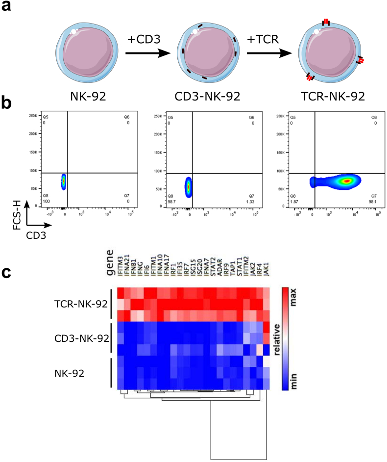
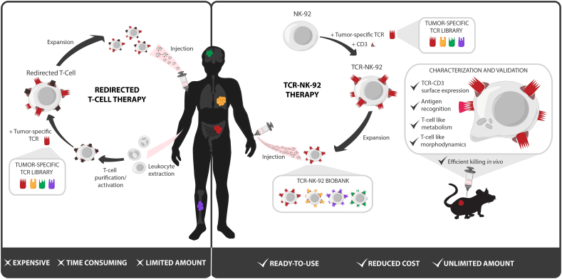
Background: Adoptive T-cell transfer of therapeutic TCR holds great promise to specifically kill cancer cells, but relies on modifying the patient's own T cells ex vivo before injection. The manufacturing of T cells in a tailor-made setting is a long and expensive process which could be resolved by the use of universal cells. Currently, only the Natural Killer (NK) cell line NK-92 is FDA approved for universal use. In order to expand their recognition ability, they were equipped with Chimeric Antigen Receptors (CARs). However, unlike CARs, T-cell receptors (TCRs) can recognize all cellular proteins, which expand NK-92 recognition to the whole proteome.

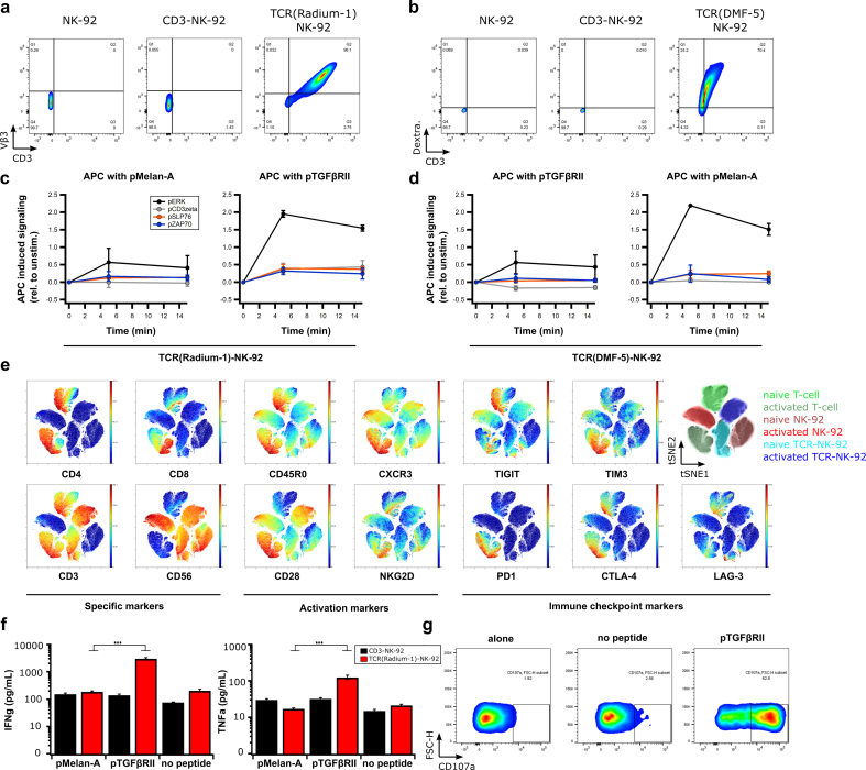
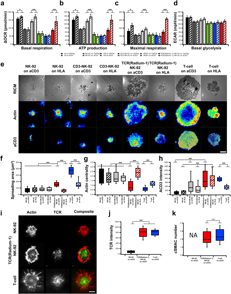
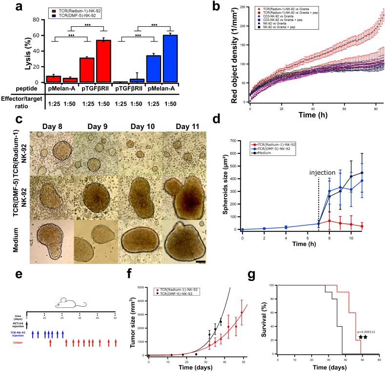
Methods: We herein genetically engineered NK-92 to express the CD3 signaling complex, and showed that it rendered them able to express a functional TCR. Functional assays and in vivo efficacy were used to validate these cells.

Findings: This is the first demonstration that a non-T cell can exploit TCRs. This TCR-redirected cell line, termed TCR-NK-92, mimicked primary T cells phenotypically, metabolically and functionally, but retained its NK cell effector functions. Our results demonstrate a unique manner to indefinitely produce TCR-redirected lymphocytes at lower cost and with similar therapeutic efficacy as redirected T cells.

Interpretation: These results suggest that an NK cell line could be the basis for an off-the-shelf TCR-based cancer immunotherapy solution. FUND: This work was supported by the Research Council of Norway (#254817), South-Eastern Norway Regional Health Authority (#14/00500-79), by OUS-Radiumhospitalet (Gene Therapy program) and the department of Oncology at the University of Lausanne.

Keywords: Immunotherapy; Natural killer; T cell; TCR.


Figures

Fig. 1
Fig. 1
Generation of a universal T cell-like lymphocyte for TCR expression. (a) Schematic overview of engineered TCR-NK-92 cells. NK-92 cells were retrovirally transduced with the human CD3 polycistronic complex, CD3εγ, CD3εδ, and CD3ζζ dimers. CD3 engineered NK-92 cells were herein referred to as CD3-NK-92. The CD3 complex is localized in the intracellular compartment. CD3-NK-92 were then super-infected with a TCRα-2A-TCRβ construct encoding a therapeutic TCR and referred to as TCR-NK-92. The CD3-TCR complex is translocated on the cell surface. (b) Dot plots showing surface expression of CD3 in TCR-NK-92 (Radium-1 or DMF-5), CD3-NK-92 and parental NK-92 (representative of three experiments). (c) Heatmap showing the relative expression of a relevant subset of genes belonging to the Interferon-signaling pathway for three replicates of TCR-NK-92, CD3-NK-92 and NK-92 in a pair-wise distance based on RNA sequencing.
Fig. 2
Fig. 2
TCR-NK-92 in vitro validation. (a,b) Dot plots showing correct folding of two transgenic TCRs (Radium-1 TCR and DMF-5 TCR). Surface expression of aCD3 and aVb3 TCR antibodies in NK-92, CD3-NK-92 and TCR(Radium-1)-NK-92 (left panel) and expression of aCD3 antibody and pMHC dextramer in NK-92, CD3-NK-92 and TCR(DMF-5)-NK-92 (right panel) (representative of three experiments). (c,d) Phosphorylation of the key components of TCR signaling cascade, ERK, CD3zeta, SLP76 and ZAP70 overtime in TCR-NK-92 (either DMF-5 TCR or Radium-1 TCR) was detected by phospho-specific flow cytometry upon stimulation with APCs loaded with either pMelan-A or pTGFβRII peptide. TCR-induced phosphorylation was measured at 5, 15 min after addition of APCs to TCR-NK-92 cells, following a 30 s centrifugation to bring the cells together. (n = 2) Error bars represent SD. (e) Outcome of ViSNE analysis on TCR-NK-92, NK-92 and T cells, either naïve or activated. (f) Bar graphs showing the level of IFNγ and TNFα produced by TCR(Radium-1)-NK-92 and CD3-NK-92 co-cultured with K562:HLA-A2 that were loaded with either irrelevant peptide (pMelan-A) or cognate peptide (pTGFβRII) or used as negative control (no peptide). (n = 3). Error bars represent SEM. (g) Dot plots showing frequency of activated TCR(Radium-1)-NK-92 cells. Detection of CD107a expression in TCR-NK-92 cells cultured in the presence of cognate peptide (pTGFβRII). (n = 4). Error bars represent SEM.
Fig. 3
Fig. 3
TCR-NK-92 acquire T-cell like behaviors in terms of metabolic functions, morphodynamics and immune synapse. (a-c) Mitochondrial respiration. Oxygen Consumption Rate (OCR) of NK-92, CD3-NK-92, TCR(Radium-1)-NK-92, J6, J76 and J Radium-1 over time, in the presence or not of anti-CD3 (aCD3). Mitochondrial respiration function (expressed as OCR rate) was measured, first at baseline and, then, probed by the serial addition of oligomycin, FCCP and antimycin-A/rotenone (anti-A + rot). (a) Basal respiration, (b) ATP production and (c) maximal respiration capacity measured in NK-92, CD3-NK-92, TCR(Radium-1)-NK-92, J6, J76 and J Radium-1, either CD3 treated or untreated. (n = 21). Error bars represent SD. (d) ExtraCellular Acidification Rate (ECAR) of NK-92, CD3-NK-92, TCR(Radium-1)-NK-92, J6, J76 and J Radium-1, either CD3 treated or untreated, before and after addition of oligomycin is shown. Comparison of basal glycolytic activity between effector cells, either CD3 treated or untreated. (n = 21). Error bars represent SD. (e) Representative Reflection Interference Contrast (RIC) and TIRF micrographs of lymphocyte adhesion (RICM), actin and CD3 organization obtained after 20 min of spreading on glass slides coated with anti-HLA class I or -CD3. Scale bar 5 μm. (f) Box plot of contact area as determined from segmentation of RICM images. (g) Box plot of actin centrality. (h) Box plot of CD3 intensity at the cell-surface contact zone. For all box plots, n ≥ 50 cells per condition. (i) Confocal images of NK-92, TCR-NK-92 and T-cell previously activated and marked with anti Vβ3 (green) and phalloidin (red). Scale bar 4 μm. (j) Box plot of the anti Vβ3 signal intensity (n = 25). Error bars represent SD. (k) Box plot of the centralization of TCR molecules (cSMAC-number) (n = 25). Error bars represent SD. (For interpretation of the references to color in this figure legend, the reader is referred to the web version of this article.)
Fig. 4
Fig. 4
TCR-NK-92 mimic T cells in their lytic effector functions both in vitro and in vivo. (a)-Antigen-specific lytic function of TCR-NK-92 cells (Radium-1 and DMF-5) measured by europium release assay. Bar graph showing % of target cells (K562:HLA-A2), either loaded or not with the cognate peptide, that were killed at 50:1 or 25:1 E:T ratio. (n = 3). Error bars represent SD. (b) Kinetic of tumor cell killing. TCR(Radium-1)-NK-92, NK-92 and CD3-NK-92 were co-cultured with Granta-519 cells either expressing or not the cognate peptide (pep). The killing is expressed as red object density by Annexin V assay. (n = 32). Error bars represent SD. (c) Representative micrographs of HCT116 spheroids treated with TCR(Radium-1)-NK-92, TCR(DMF-5)-NK-92 or medium. Scale bar 200 μm. (d) Evolution of the average spheroids size upon treatment with TCR(Radium-1)-NK-92, TCR(DMF-5)-NK-92 or medium. (n = 28). Error bars represent SD. (e) Design of in vivo experiment using HCT116 xenograft mouse model. 10 mice per condition. (f) Evolution of tumor volume over time in treated (TCR(Radium-1)-NK-92) and untreated (TCR(DMF-5)-NK-92) mice. Error bars represent SD. (g) Corresponding Kaplan-Meier estimator. (For interpretation of the references to color in this figure legend, the reader is referred to the web version of this article.)
Fig. 5
Fig. 5
Technical advantages of TCR-NK-92 based immunotherapy over T-cell based immunotherapy.

References

    1. Fesnak A.D., June C.H., Levine B.L. Engineered T cells: the promise and challenges of cancer immunotherapy. Nat Rev Cancer. 2016;16:566–581. - PMC - PubMed
    1. Johnson L.A., June C.H. Driving gene-engineered T cell immunotherapy of cancer. Cell Res. 2017;27:38–58. - PMC - PubMed
    1. Rosenberg S.A., Restifo N.P., Yang J.C., Morgan R.A., Dudley M.E. Adoptive cell transfer: a clinical path to effective cancer immunotherapy. Nat Rev Cancer. 2008;8:299–308. - PMC - PubMed
    1. Rosenberg S.A., Restifo N.P. Adoptive cell transfer as personalized immunotherapy for human cancer. Science. 2015;348:62–68. (80- ) - PMC - PubMed
    1. Themeli M., Rivière I., Sadelain M. New Cell sources for T Cell Engineering and Adoptive Immunotherapy. Cell Stem Cell. 2015;16:357–366. - PMC - PubMed

MeSH terms