Review
. 2019 Jan 7:6:170.
doi: 10.3389/fcell.2018.00170. eCollection 2018.

Wnt Pathway in Bone Repair and Regeneration - What Do We Know So Far

Affiliations
Review

Wnt Pathway in Bone Repair and Regeneration - What Do We Know So Far

Khosrow S Houschyar et al. Front Cell Dev Biol. .

Abstract

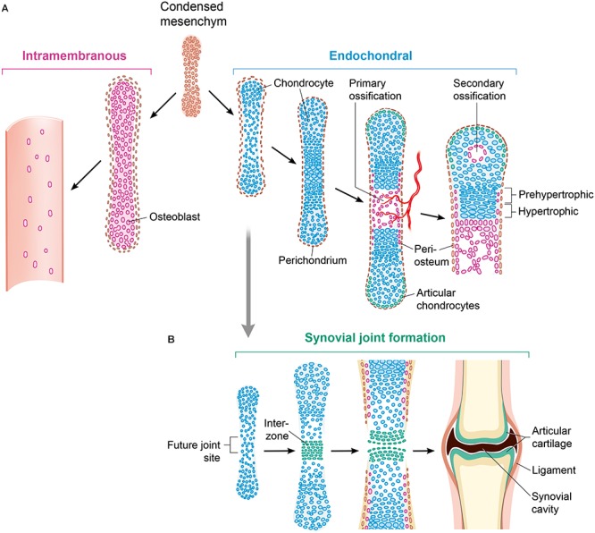
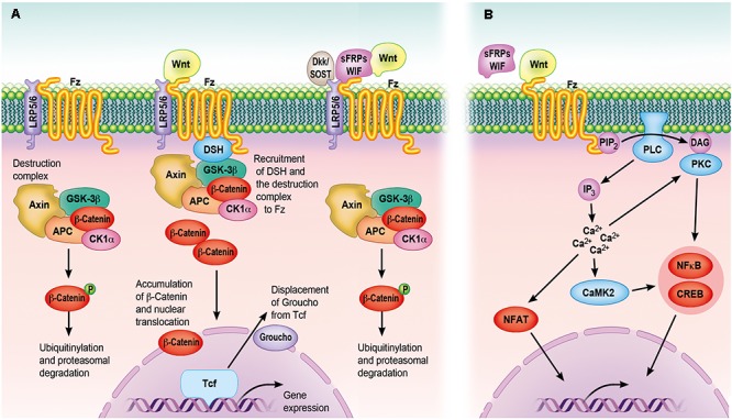
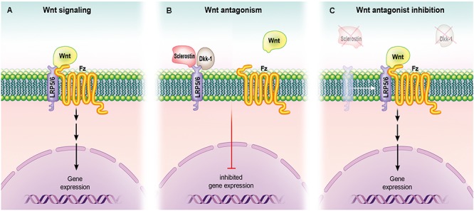
Wnt signaling plays a central regulatory role across a remarkably diverse range of functions during embryonic development, including those involved in the formation of bone and cartilage. Wnt signaling continues to play a critical role in adult osteogenic differentiation of mesenchymal stem cells. Disruptions in this highly-conserved and complex system leads to various pathological conditions, including impaired bone healing, autoimmune diseases and malignant degeneration. For reconstructive surgeons, critically sized skeletal defects represent a major challenge. These are frequently associated with significant morbidity in both the recipient and donor sites. The Wnt pathway is an attractive therapeutic target with the potential to directly modulate stem cells responsible for skeletal tissue regeneration and promote bone growth, suggesting that Wnt factors could be used to promote bone healing after trauma. This review summarizes our current understanding of the essential role of the Wnt pathway in bone regeneration and repair.

Keywords: Wnt; bone; canonical; non-canonical; regeneration; repair; stem cells; β-catenin.


Figures

FIGURE 1
FIGURE 1
Ways of bone formation. (A) Ossification can occur via endochondral or intramembranous mechanisms. As part of the intramembranous ossification, mesenchymal cells differentiate directly into osteoblasts and generate bone tissue. Chondrocytes develop from mesenchymal cell differentiation with forming an intermediate cartilage during endochondral ossification. By mineralizing the matrix, undergoing apoptosis and attracting blood vessels and osteoblasts, hypertrophying chondrocytes that stop proliferating initiate a centric growth plate. (B) Histologically detectable flattening and gathering of cells that are forming an interzone, is the first sign of joint formation. This is followed by maturation and remodeling leading to a mature synovial joint. The Wnt signaling pathway is crucial for controlling almost all aspects of this skeleton formation. Osteoblasts (purple); chondrocytes (blue); osteochondroprogenitor cells (brown).
FIGURE 2
FIGURE 2
The Wnt signaling cascades. (A) The canonical Wnt signaling cascade depends on β-catenin, which serves as an intracellular signaling molecule. In case Wnt is not binding to Fz receptors, β-catenin is sequestered into a destruction complex composed of Axin, CK1α, APC and GSK3β, phosphorylated, ubiquitinylated and subsequently degraded by the proteasome. Following the binding of Wnt to Fz receptors and LRP5/6 co-receptors, DSH recruits the destruction complex to the cell membrane by interacting with the receptor complex. This allows newly synthesized β-catenin to accumulate within the cytoplasm and to translocate to the nucleus. By displacing the transcriptional co-repressor groucho from TCF transcription factors, nuclear β-catenin can activate a gene transcription program, whereas Wnt-binding antagonists (sFRPs/WIF) and Wnt receptor antagonists (Dkk/SOST) inhibit the canonical cascade. (B) The non-canonical Wnt signaling cascade is characterized by the activation through phosphorylation cascades, which are themselves activated by specific ligand–receptor interactions, seemingly without engagement of the LRP co-receptors. Increasing intracellular Ca2+ levels following PLC- and DAG production can trigger many of these cascades. Subsequently, PKC and CaMKII can activate transcription factors like NFκB and CREB, mediated IP3 and calmodulin is involved in the activation of NFAT. However, only the Wnt-binding antagonists are able to inhibit the non-canonical cascade. APC, adenomatous polyposis coli; CaMKII, calcium/calmodulin-dependent protein kinase type II; CK1α, caseine kinase 1-α; CREB, cyclic AMP-responsive element-binding protein; DAG, diacylglycerol; Dkk, Dickkopf; DSH, disheveled; GSK3β, glycogen synthase kinase-3 β; IP3, inositol 1,4,5-triphosphate; LRP, low-density lipoprotein receptor-related protein; NFAT, nuclear factor of activated T cells; NFκB, nuclear factor κB; PIP2, phosphatidylinositol 4,5-bisphosphate; PKC, protein kinase C; PLC, phospholipase C; sFRPs, secreted frizzled-related proteins; SOST, sclerostin; WIF, Wnt inhibitory factor.
FIGURE 3
FIGURE 3
Role of Wnt signaling in osteoblasts. (A) Upon binding to its receptor (Frizzled) and co-receptors (LRP5 and LRP6), Wnt activates their signaling pathway, leading to gene expression (and ultimately protein synthesis and the formation of bone). (B) Wnt antagonists sclerostin and Dkk-1 bind LRP5 and LRP6, preventing their interaction with Frizzled and resulting in inhibition of gene expression. (C) Loss-of-function mutation in a gene that encodes for a Wnt antagonist orpharmacological engagement of the antagonist with an inhibitory molecule such as an antibody can lead to inhibition of Wnt antagonism and promote gene expression.

Similar articles

Cited by

References

    1. Amini A. R., Laurencin C. T., Nukavarapu S. P. (2012). Bone tissue engineering: recent advances and challenges. Crit. Rev. Biomed. Eng. 40 363–408. 10.1615/CritRevBiomedEng.v40.i5.10 - DOI - PMC - PubMed
    1. Arnsdorf E. J., Tummala P., Jacobs C. R. (2009). Non-canonical Wnt signaling and N-cadherin related beta-catenin signaling play a role in mechanically induced osteogenic cell fate. PLoS One 4:e5388. 10.1371/journal.pone.0005388 - DOI - PMC - PubMed
    1. Arvidson K., Abdallah B. M., Applegate L. A., Baldini N., Cenni E., Gomez-Barrena E., et al. (2011). Bone regeneration and stem cells. J. Cell. Mol. Med. 15 718–746. 10.1111/j.1582-4934.2010.01224.x - DOI - PMC - PubMed
    1. Balemans W., Van Hul W. (2007). The genetics of low-density lipoprotein receptor-related protein 5 in bone: a story of extremes. Endocrinology 148 2622–2629. 10.1210/en.2006-1352 - DOI - PubMed
    1. Bao Q., Chen S., Qin H., Feng J., Liu H., Liu D., et al. (2017). An appropriate Wnt/beta-catenin expression level during the remodeling phase is required for improved bone fracture healing in mice. Sci. Rep. 7:2695. 10.1038/s41598-017-02705-0 - DOI - PMC - PubMed

LinkOut - more resources