. 2019 Jun;20(3):303-319.
doi: 10.1007/s10522-018-09792-0. Epub 2019 Jan 21.

Generation of a novel model of primary human cell senescence through Tenovin-6 mediated inhibition of sirtuins

Affiliations

Generation of a novel model of primary human cell senescence through Tenovin-6 mediated inhibition of sirtuins

Hannah E Walters et al. Biogerontology. 2019 Jun.

Abstract

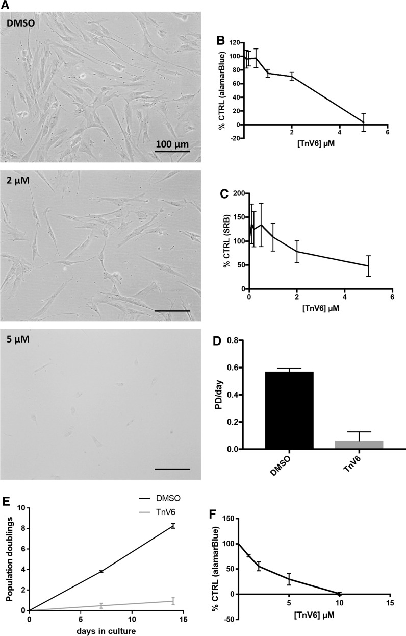
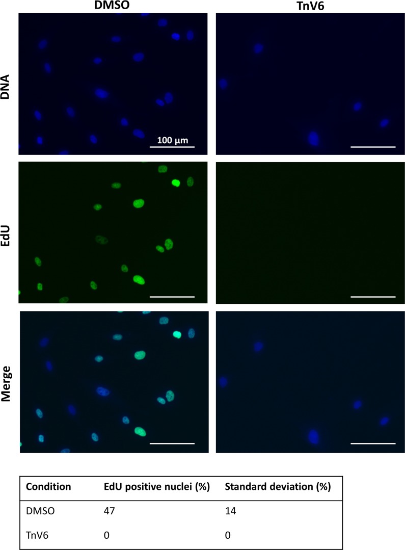
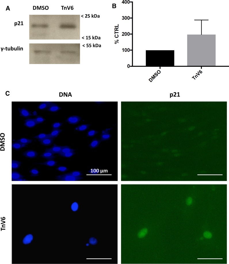
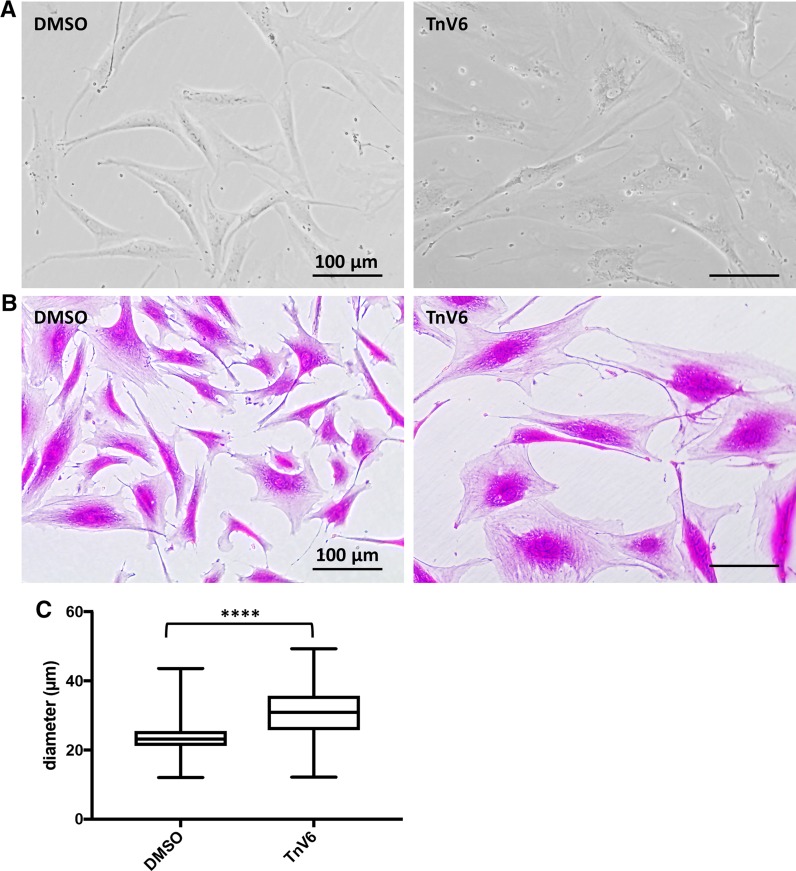
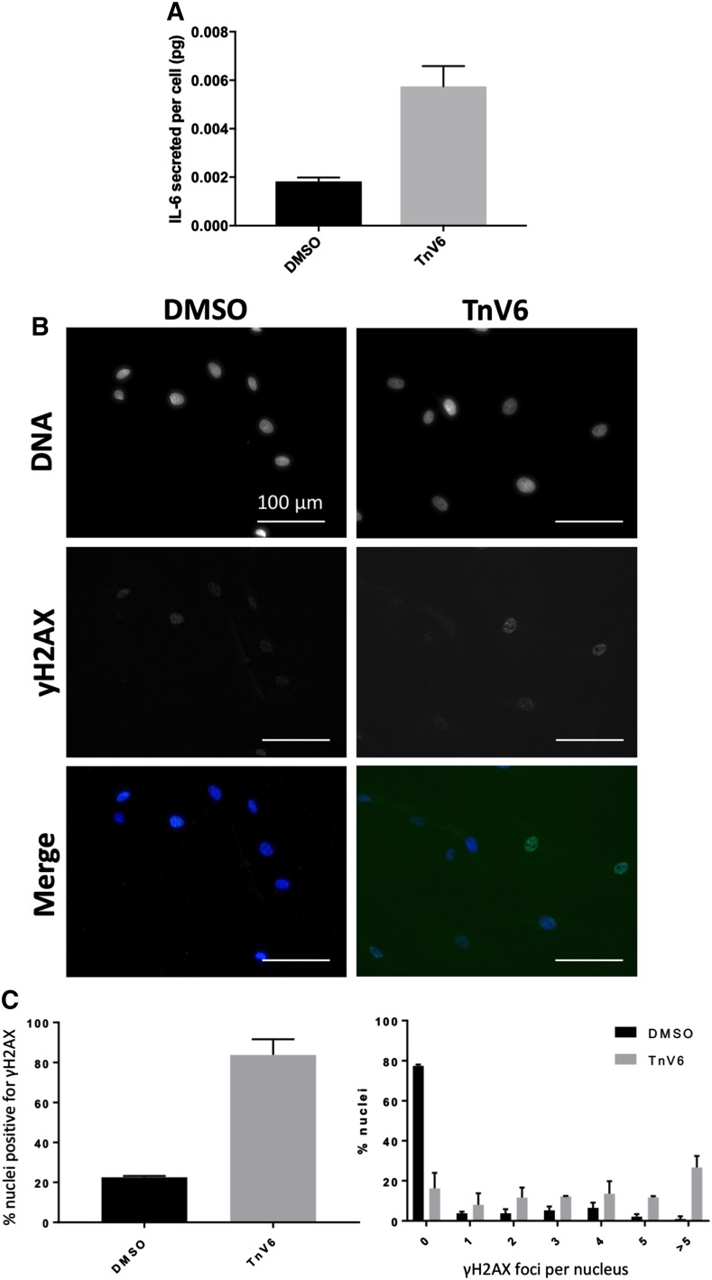
Cell senescence, a state of cell cycle arrest and altered metabolism with enhanced pro-inflammatory secretion, underlies at least some aspects of organismal ageing. The sirtuin family of deacetylases has been implicated in preventing premature ageing; sirtuin overexpression or resveratrol-mediated activation of sirtuins increase longevity. Here we show that sirtuin inhibition by short-term, low-dose treatment with the experimental anti-cancer agent Tenovin-6 (TnV6) induces cellular senescence in primary human fibroblasts. Treated cells cease proliferation and arrest in G1 of the cell cycle, with elevated p21 levels, DNA damage foci, high mitochondrial and lysosomal load and increased senescence-associated β galactosidase activity, together with actin stress fibres and secretion of IL-6 (indicative of SASP upregulation). Consistent with a histone deacetylation role of SIRT1, we find nuclear enlargement, possibly resulting from chromatin decompaction on sirtuin inhibition. These findings highlight TnV6 as a drug that may be useful in clinical settings where acute induction of cell senescence would be beneficial, but also provide the caveat that even supposedly non-genotoxic anticancer drugs can have unexpected and efficacy-limiting impacts on non-transformed cells.

Keywords: Ageing; HDAC/KDAC; Longevity; SASP; Senescence; Sirtuin; Tenovin-6; p21.


Conflict of interest statement

The authors declare no conflicting financial interests.

Figures

Fig. 1
Fig. 1
TnV6 strongly suppresses HDAC activity in both primary and cancer cells. Inhibition of deacetylase activity was measured using the Fluor de Lys® HDAC fluorometric cellular activity assay (deacteylation of a substrate to generate a fluorescent product) on HeLa or HF043 cells plated in triplicate wells of 96 well plates. Cells were treated with DMSO (vehicle control), resveratrol (RSV, 50 µM), trichostatin A (TSA, 1 µM) or TnV6 (2 µM). HF043 and HeLa experiments were performed on different days (n = 2, data from one representative experiment per cell line shown; statistical analysis in Supplementary Table S1)
Fig. 2
Fig. 2
Low dose TnV6 halts proliferation in primary human fibroblasts. a Phase contrast microscopy images of HF043 fibroblasts (at low cumulative population doubling < 50) treated with DMSO, 2 µM or 5 µM Tnv6 for 24 h (n > 3). b Analysis of reducing capacity, taken as a proxy for cell viability, using the alamarBlue vital dye, following treatment of HF043 fibroblasts with TnV6 concentrations ranging from 0.1 µM to 5 µM for 72 h (n = 3, mean ± SD). c Total cell biomass was measured by staining with sulforhodamine B (SRB) on cells treated as in (b) (n = 3, mean ± SD). d Proliferation rate measured as population doublings (PD) per day for HF043 fibroblasts cultured with either DMSO or 2 µM TnV6 for 7 days. Cell counting was performed using a Cellometer T4 (n = 3). 2-tailed unpaired t-test *** p = 0.0002. e Number of population doublings over a 14-day period was measured, as in (d), with control cells treated with DMSO throughout, and TnV6 cells treated with 2 µM TnV6 for the first 7 days, and DMSO for days 7–14. f HeLa cells were treated with a range of concentrations of TnV6 for 72 h before viability analysis using alamarBlue
Fig. 3
Fig. 3
Ablation of DNA replication in primary human fibroblasts on low dose TnV6 treatment. DNA replication was assessed in HF043 fibroblasts treated for 72 h with either DMSO or 2 µM TnV6, before incubation with 10 µM EdU for 18 h, fixation and staining using Click-IT chemistry for incorporated EdU. Cells were then imaged on a ZOETM fluorescence imager with gain and contrast settings kept identical between wells and images. Fiji software was used for quantification. Technical triplicates were conducted for each experiment, with at least 50 cells analysed per well; number of biological replicates n = 3, 2-tailed unpaired t-test ** p = 0.0043
Fig. 4
Fig. 4
G1 phase arrest in primary human fibroblasts treated with TnV6. Cell cycle stage was assessed in HF043 fibroblasts treated with DMSO or 2 µM Tnv6 in triplicate for 72 h using the Cell Cycle Clock dye reagent. Images of stained cells were then analysed by Fiji (n = 3, > 50 cells per well analysed, data from one representative experiment shown). * p < 0.05
Fig. 5
Fig. 5
TnV6 induces p21 upregulation in primary human fibroblasts. a Western blot of lysates of HF043 fibroblasts treated with either DMSO (control) or 2 µM TnV6 for 7 days, probed with anti-p21 antibody (upper panel) or anti- γ-tubulin antibody (lower panel). Marker sizes shown by arrows on right. b Bands on the blots were quantified by densitometry using Fiji software and then the p21 signal was normalised against the tubulin loading control (n = 3). c Immunofluorescence of HF043 fibroblasts treated with DMSO or 2 µm TnV6 for 7 days then probed with anti-p21 antibody and Alexafluor-488 secondary antibody, with DNA counterstained with NucBlue live
Fig. 6
Fig. 6
Extended TnV6 treatment induces senescent morphology. a Phase contrast microscopy of HF043 fibroblasts treated with DMSO or 2 µM TnV6 for 7 days (n > 3). b Cells as in a were fixed and stained with sulforhodamine B then imaged using transmission light microscopy (n > 3). c Cell diameter was analysed for cells treated as above, with > 30 cells counted per sample (n > 3 biological replicates, results from one representative experiment shown). Box shows 25th and 75th percentiles with median line; bars show maximum and minimum, **** p < 0.001
Fig. 7
Fig. 7
Senescence phenotypes result from long term TnV6 exposure. a SAβGAL staining of HF043 fibroblasts treated for 7 days with DMSO or 2 µM TnV6. b Lysosomal analysis by Lysotracker Red staining of HF043 fibroblasts treated for 72 h with DMSO or 2 µM TnV6; DNA was counterstained with NucBlue Live. Images were acquired in living cells on a ZOE fluorescence imager with gain and contrast settings constant throughout. c Nuclear area was analysed from fluorescence microscopy images of cells stained with NucBlue Live and quantified by Fiji (n > 3, > 50 nuclei counted per sample, data from one representative experiment shown). **** p < 0.001. d Mitochondrial staining of HF043 fibroblasts after 7-day treatment with DMSO or 2 µM TnV6 using Mitotracker Green and imaged live (n = 2). e Rhodamine-phalloidin staining for actin in HF043 fibroblasts fixed and stained after 7-day treatment with DMSO or 2 µM TnV6. DNA was counterstained with NucBlue Live (n = 2)
Fig. 8
Fig. 8
IL-6 is upregulated in TnV6-treated cells and associates with elevation of DNA damage. a ELISA was used to measure IL-6 secreted into 24-hour conditioned medium by HF043 fibroblasts treated with 2 µM TnV6 or DMSO for 7 days in total. Recombinant human IL-6 was used to generate a standard curve for determination of IL-6 amounts, and IL-6/cell (pg) was calculated following cell counting. n = 3, mean ± S.D. are shown, 2-tailed unpaired t-test p = 0.0059. b Analysis of DNA damage by immunofluorescence for γH2AX in HF043 fibroblasts treated for 7 days with 2 µM TnV6 or DMSO, with DNA counterstained by NucBlue Live. c Quantitation of  % nuclei with γH2AX staining (2-tailed unpaired t-test p = 0.0002) and number of γH2AX foci per nucleus. Mean ± S.D. shown

Similar articles

Cited by

References

    1. Aparicio OM, Billington BL, Gottschling DE. Modifiers of position effect are shared between telomeric and silent mating-type loci in S. cerevisiae. Cell. 1991;66:1279–1287. doi: 10.1016/0092-8674(91)90049-5. - DOI - PubMed
    1. Baar MP, et al. Targeted apoptosis of senescent cells restores tissue homeostasis in response to chemotoxicity and aging. Cell. 2017;169(132–147):e116. - PMC - PubMed
    1. Baker DJ, et al. Naturally occurring p16(Ink4a)-positive cells shorten healthy lifespan. Nature. 2016;530:184–189. doi: 10.1038/nature16932. - DOI - PMC - PubMed
    1. Bridger JM, Boyle S, Kill IR, Bickmore WA. Re-modelling of nuclear architecture in quiescent and senescent human fibroblasts. Curr Biol. 2000;10:149–152. doi: 10.1016/S0960-9822(00)00312-2. - DOI - PubMed
    1. Burnett C, et al. Absence of effects of Sir2 overexpression on lifespan in C. elegans and Drosophila. Nature. 2011;477:482–485. doi: 10.1038/nature10296. - DOI - PMC - PubMed

Publication types

LinkOut - more resources