. 2019 Jun;8(6):575-585.
doi: 10.1002/sctm.18-0141. Epub 2019 Jan 21.

Chondroitin Sulfate Glycosaminoglycan Scaffolds for Cell and Recombinant Protein-Based Bone Regeneration

Affiliations

Chondroitin Sulfate Glycosaminoglycan Scaffolds for Cell and Recombinant Protein-Based Bone Regeneration

Seth Andrews et al. Stem Cells Transl Med. 2019 Jun.

Abstract

Bone morphogenetic protein 2 (BMP-2)-loaded collagen sponges remain the clinical standard for treatment of large bone defects when there is insufficient autograft, despite associated complications. Recent efforts to negate comorbidities have included biomaterials and gene therapy approaches to extend the duration of BMP-2 release and activity. In this study, we compared the collagen sponge clinical standard to chondroitin sulfate glycosaminoglycan (CS-GAG) scaffolds as a delivery vehicle for recombinant human BMP-2 (rhBMP-2) and rhBMP-2 expression via human BMP-2 gene inserted into mesenchymal stem cells (BMP-2 MSC). We demonstrated extended release of rhBMP-2 from CS-GAG scaffolds compared to their collagen sponge counterparts, and further extended release from CS-GAG gels seeded with BMP-2 MSC. When used to treat a challenging critically sized femoral defect model in rats, both rhBMP-2 and BMP-2 MSC in CS-GAG induced comparable bone formation to the rhBMP-2 in collagen sponge, as measured by bone volume, strength, and stiffness. We conclude that CS-GAG scaffolds are a promising delivery vehicle for controlling the release of rhBMP-2 and to mediate the repair of critically sized segmental bone defects. Stem Cells Translational Medicine 2019;8:575-585.

Keywords: Bone morphogenetic protein-2; Chondroitin sulfate glycosaminoglycan; Genetic therapy; Mesenchymal stromal cells; Osteogenesis.


Conflict of interest statement

The authors indicated no potential conflicts of interest.

Figures

Figure 1
Figure 1
Efficient transduction of mesenchymal stem cells with a lentiviral vector at 10 MOI. (A): Images of uMSC over 72 hours after transducing with prEF1a‐RFP at 10 and 50 MOI under phase contrast and RFP filter to evaluate transduction efficiency. Scale bars indicate 200 μm. N = 15. (B): Mean BMP‐2 expression in uMSCs and bmMSCs at 48, 72, 96, and 120 hours after transducing with prEF1a‐BMP2 at 10 and 50 MOI, comparing expression over cell type and MOI within each time point. Transductions at 0 MOI did not result in BMP‐2 expression detectable by BMP‐2 enzyme‐linked immunosorbent assay. Groups with differing letters are significantly different from each other within a time point at p < .05. Error bars indicate standard error. Two‐way analysis of variance with Tukey's multiple comparison test (n = 5–6). Abbreviations: bmMSC, bone marrow mesenchymal stem cell; BMP‐2, bone morphogenetic protein 2; MOI, moment of inertia; uMSC, umbilical mesenchymal stem cell.
Figure 2
Figure 2
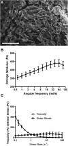
Chondroitin sulfate glycosaminoglycan (CS‐GAG) hydrogel is porous with stable rheology. (A): Scanning electron microscopy of CS gel surface at ×165 magnification. Pore sizes are approximately 20–100 μm. Scale bar indicates 500 μm. (B): Rheology of CS‐GAG gel. Data indicate stable rheological properties across a range of frequencies. (C): Dynamic viscosity decreases, and shear stress increases with an increase in shear rate. Error bars indicate standard error (n = 5).
Figure 3
Figure 3
Preparation of chondroitin sulfate GAG hydrogel and interactions with transduced MSCs. (A): Casting of 3% GAG hydrogel (i) followed by photo crosslinking under UV light (ii). The hydrogel is lyophilized for 24 hours (iii) before it is rehydrated with a cell suspension in basal media (iv). (B): Measures of colocalization for calcein and RFP: Pearson's correlation, describing the extent of overlap between RFP and calcein images, and colocalization coefficients M1 and M2, describing the fraction of RFP colocalizing with calcein and the fraction of calcein localizing with RFP, respectively (n = 5). (C): Cumulative BMP‐2 release over 15 days from empty GAG gels, GAG gels loaded with 1 million nontransduced MSCs, and GAG gels loaded with 1 million BMP‐2 MSCs (n = 3–4). (D): BMP‐2 release kinetics shown as percentage of the total amount released for GAG gels loaded with BMP‐2 MSCs, as well as GAG gels and collagen sponges loaded with rhBMP‐2 (n = 3–4). Abbreviations: BMP‐2, bone morphogenetic protein 2; GAG, glycosaminoglycan; MSC, mesenchymal stem cell; rhBMP‐2, recombinant human BMP‐2.
Figure 4
Figure 4
Defects bridge when treated with rhBMP‐2 or BMP‐2 MSCs. (A): Representative longitudinal radiographs at 4 and 12 weeks postsurgery. Defects were treated with 1 million nontransduced umbilical mesenchymal stem cells (uMSCs) in 150 μl chondroitin sulfate (CS)‐GAG gel, 1 million BMP‐2 uMSCs in 150 μl CS‐GAG gel, 150 μl CS‐GAG hydrogel loaded with 5 μg rhBMP‐2, or 150 μL collagen sponge loaded with 5 μg rhBMP‐2. (B): Twelve‐week micro‐computed tomography reconstructions of the same bone defects shown in the radiographs. Abbreviations: 3D, three dimension; BMP‐2, bone morphogenetic protein 2; GAG, glycosaminoglycan; MSC, mesenchymal stem cell; rhBMP‐2, recombinant human BMP‐2.
Figure 5
Figure 5
Newly formed bone similar between rhBMP‐2 and BMP‐2 MSC groups. Micro‐computed tomography characterization and mechanical testing of regenerated femurs at 12 weeks. (A): Quantification of new bone revealed GAG + BMP‐2 MSC, GAG + rhBMP‐2, and Col + rhBMP‐2 all demonstrated greater total BVs than GAG+MSC. (B): Calculated average pMOI showed that both GAG + BMP‐2 MSC and GAG + rhBMP‐2 groups had significantly higher pMOI compared to GAG + MSC. (C): Torque to failure. (D): Torsional stiffness measured from testing regenerated femurs to failure at 12 weeks. Both GAG + rhBMP‐2 and Col + rhBMP‐2 groups had significantly higher torque to failure and torsional stiffness compared to GAG + MSC. There were no significant differences among GAG + BMP‐2 MSC, GAG + rhBMP‐2, and Col + rhBMP‐2 for any of these metrics. Error bars indicate standard error (n = 7–8 per group). Nonparametric Kruskal‐Wallis test with multiple comparisons made by Dunn's post‐tests. Abbreviations: BV, bone volume; BMP‐2, bone morphogenetic protein 2; Col, collagen; GAG, glycosaminoglycan; MSC, mesenchymal stem cell; pMOI, polar moment of inertia; rhBMP‐2, recombinant human BMP‐2.
Figure 6
Figure 6
Histology reveals qualitative differences in bone maturity. Representative H&E images of defect tissue at 12 weeks postsurgery. (A): All BMP‐2 groups exhibited islands of new bone formation while GAG + MSC did not. New bone is denoted by the black arrows in the higher magnification inset. (B): Furthermore, upon inspection under polarized light to assess collagen alignment, the bone in the Col + rhBMP‐2 group appeared to be predominantly lamellar in structure, whereas both GAG + BMP‐2 MSC and GAG + rhBMP‐2 had material resembling woven bone. All images were taken in the middle of the bone defect. Abbreviations: BMP‐2, bone morphogenetic protein 2; Col, collagen; GAG, glycosaminoglycan; MSC, mesenchymal stem cell; rhBMP‐2, recombinant human BMP‐2.

References

    1. Greenwald AS, Boden SD, Goldberg VM et al. Bone‐graft substitutes: Facts, fictions, and applications. J Bone Joint Surg Am 2001, 83‐A (suppl 2; Pt 2):98–103. - PubMed
    1. Toolan BC. Current concepts review: Orthobiologics. Foot Ankle Int. 2006;27(7):561–566. - PubMed
    1. De Long WG, Einhorn TA, Koval K et al. Bone grafts and bone graft substitutes in orthopaedic trauma surgery: A critical analysis. J Bone Joint Surg Am 2007;89:649–658. - PubMed
    1. Laurencin C, Khan Y, El‐Amin SF. Bone graft substitutes. Expert Rev Med Devices 2006;3:49–57. - PubMed
    1. Desai BM. Osteobiologics. Am J Orthop (Belle Mead NJ) 2007;36 (suppl 4):8–11. - PubMed

Publication types

MeSH terms