Comparative Study
. 2019 Mar;280(3):395-410.
doi: 10.1002/jmor.20952. Epub 2019 Jan 22.

Comparative analysis of avian hearts provides little evidence for variation among species with acquired endothermy

Affiliations
Comparative Study

Comparative analysis of avian hearts provides little evidence for variation among species with acquired endothermy

Jelle G H Kroneman et al. J Morphol. 2019 Mar.

Abstract

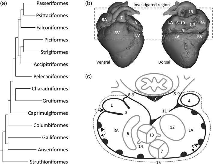
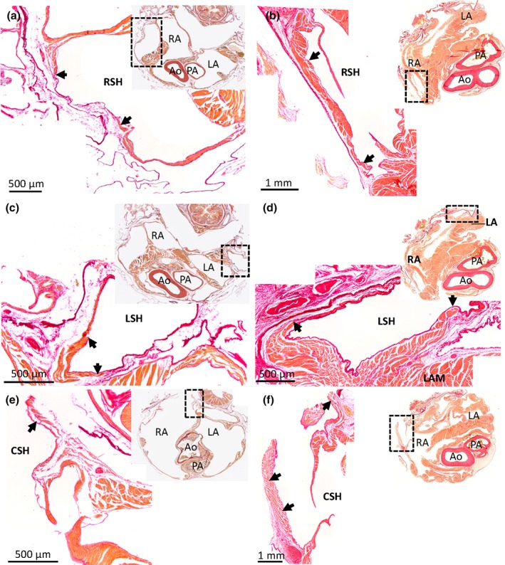
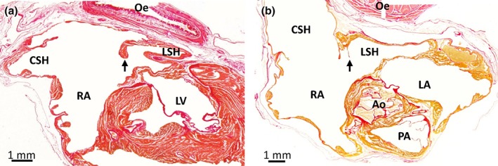
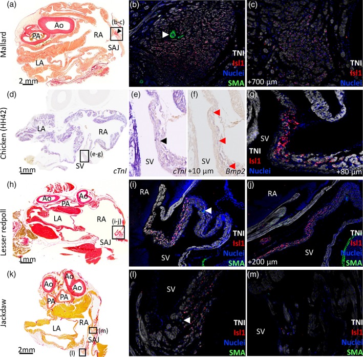
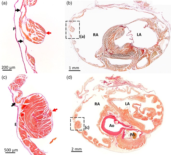
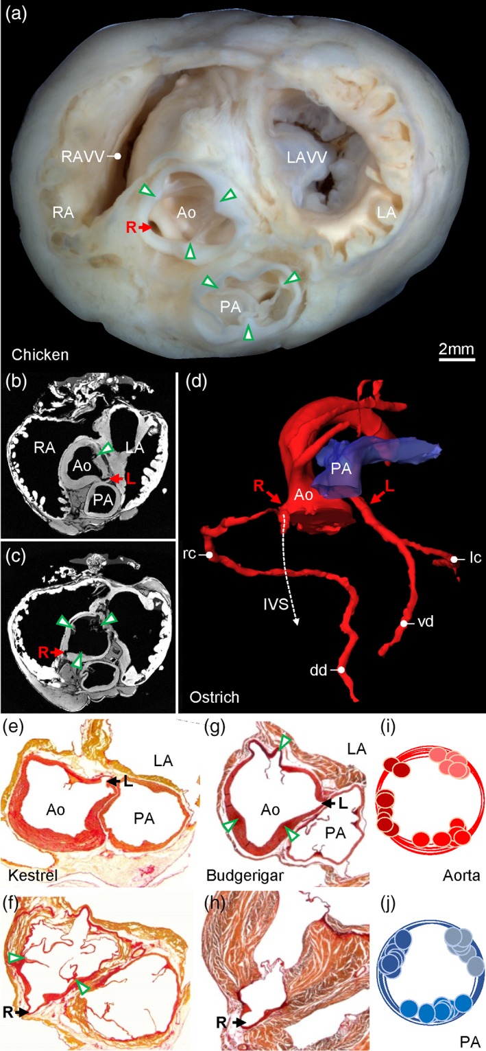
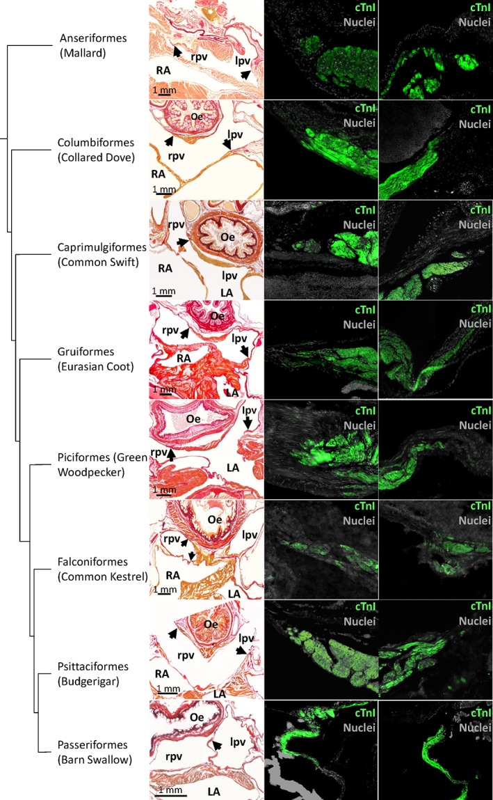
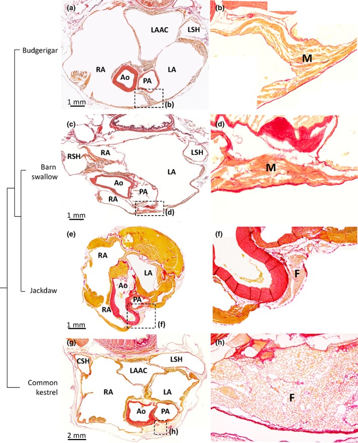
Mammals and birds acquired high performance hearts and endothermy during their independent evolution from amniotes with many sauropsid features. A literature review shows that the variation in atrial morphology is greater in mammals than in ectothermic sauropsids. We therefore hypothesized that the transition from ectothermy to endothermy was associated with greater variation in cardiac structure. We tested the hypothesis in 14 orders of birds by assessing the variation in 15 cardiac structures by macroscopic inspection and histology, with an emphasis on the atria as they have multiple features that lend themselves to quantification. We found bird hearts to have multiple features in common with ectothermic sauropsids (synapomorphies), such as the presence of three sinus horns. Convergent features were shared with crocodylians and mammals, such as the cranial offset of the left atrioventricular junction. Other convergent features, like the compact organization of the atrial walls, were shared with mammals only. Pacemaker myocardium, identified by Isl1 expression, was anatomically node-like (Mallard), thickened (Chicken), or indistinct (Lesser redpoll, Jackdaw). Some features were distinctly avian, (autapomorphies) including the presence of a left atrial antechamber and the ventral merger of the left and right atrial auricles, which was found in some species of parrots and passerines. Most features, however, exhibited little variation. For instance, there were always three systemic veins and two pulmonary veins, whereas among mammals there are 2-3 and 1-7, respectively. Our findings suggest that the transition to high cardiac performance does not necessarily lead to a greater variation in cardiac structure.

Keywords: anatomy; bird; evolution; heart.


Figures

Figure 1
Figure 1
Phylogeny and scheme of the analysed structures of the avian hearts. (a) Phylogeny, on the basis of Jarvis et al. (2014), of the investigated orders. (b) In our investigations, we focused on the left and right atrium (LA and RA, respectively), the connecting veins, and the base of the left and right ventricle (LV and RV, respectively), and the arterial bases (see below for the number coding). In subsequent figure legends, we will refer to section numbers, where Section 1 represents the top horizontal line of the dashed box. (c) Schematic section in the transverse plane, which was the principle plane of sectioning, onto which we have collapsed all investigated structures: 1, myocardium in the sinus venosus; 2, the sinuatrial node; 3, sinuatrial valve and leaflets; 4, left sinus horn; 5, pectinate muscles of the atrial wall; 6, position of the right atrioventricular junction and muscularity of the valve; 7, position of the pulmonary artery and valve leaflets; 8, number of pulmonary veins; 9, the extent of pulmonary venous myocardium relative to the pericardium (dashed line); 10, dorsal ridge of the antechamber of the left atrium; 11, muscular shelf in the roof of the left atrium; 12, position of the left atrioventricular junction and muscularity of the valve; 13, position of the aorta and valve leaflets; 14, position of the orifices of the coronary arteries; 15, presence of ventral merger of the atrial walls
Figure 2
Figure 2
Myocardium in the sinus venosus of the Budgerigar (a, c, e) and the Mallard (b, d, f). Picro‐sirius red stained 10 μm histological sections in the transverse plane. Upper‐right corner inserts show the entire section, with the black boxes indicating the magnified part. The right sinus horn (RSH) (a, b), left sinus horn (LSH) (c, d), and caudal sinus horn (CSH) (e, f) contain myocardium (black arrows). The myocardial wall can be quite thick proximal to the atria, but tapers off distally. At the pericardial border, the vessel wall may be without myocardium. In the images of the Budgerigar, blood has been painted over with white for clarity. In the Budgerigar, sections 300 (cranial) to 850 (caudal) encompassed the atria and the sections shown are 425 (a), 400 (c), 600 (e). In the Mallard, sections 271 (cranial) to 1201 (caudal) encompassed the atria and the sections shown are 511 (b), 541 (d), 691 (f). Ao = aorta; LA = left atrium; LAM = left atrial muscle; PA = pulmonary artery; RA = right atrium
Figure 3
Figure 3
Leaflet of the valve of the left sinus horn in the Green woodpecker (a) and the Common kestrel (b). Birds generally have a prominent valve (black arrow) guarding the orifice of the left sinus horn (LSH), here exemplified by the Green woodpecker (section 1101, transverse plane, is shown where sections between 701 (cranial) and 1381 (caudal) encompassed the atria). It was found on sections representing 0.7 mm out of the total height of the atria of 6.8 mm. In the Common kestrel, the valve was a very thin membranous leaflet and it was found on sections representing 0.7 mm out of the total height of the atria of 6.6 mm (section 881, transverse plane, is shown where sections between 501 (cranial) and 1281 (caudal) encompassed the atria). Ao = aorta; CSH = caudal sinus horn; Oe = oesophagus; LA = left atrium; LV = left ventricle; LSH = left sinus horn; PA = pulmonary artery; RA = right atrium. In the images, blood has been painted over with white for clarity
Figure 4
Figure 4
Sinuatrial node in Mallard (a–c), chicken HH42 (d–g), Lesser redpoll (h–j), and Jackdaw (k–m). All sections are in the transverse plane. The boxed areas in the left‐hand column images indicate the areas of the images of the middle and right‐hand columns. (a–c) In the Mallard, a nodal structure at the base of the right leaflet of the sinuatrial valve (black arrowhead in [a]) expressed Isl1 (b, c) and had a large coronary artery (white arrowhead in [b]). Sections 271 (cranial) to 1201 (caudal) encompassed the atria and the sections shown are 691 (a), 692 (b), 762 (c). (d–g) In the chicken HH42, the sinus venosus (SV) expressed the myocardial marker cTnI and this expression was relatively weak at the base of the right leaflet of the sinuatrial valve (black arrowhead in [e]). (f–g) The base of the right leaflet of the sinuatrial valve expressed Bmp2 (red arrowheads in [f]) and Isl1 [g]). Sections 17 (cranial) to 297 (caudal) encompassed the atria and the sections shown are 80 (d‐e), 78 (f), 88 (g). The sections were from the mid‐height of the atria. (h–j) In the lesser redpoll, Isl1 was expressed in the base of the right leaflet of the sinuatrial valve. There was no nodal structure but the Isl1 expressing wall was thicker than the surrounding walls and contained a large coronary artery (white arrowhead in [i]). Sections 321 (cranial) to 621 (caudal) encompassed the atria and the sections shown are 401 (h), 402 (i), 422 (j). (k–m) In the Jackdaw an Isl1 positive area was seen in the left sinus venosus myocardium (l) in which a coronary artery was visible (white arrowhead in (l). At the base of the right sinuatrial leaflet no positive Isl1 cells could be seen (m). Sections 122 (cranial) to 332 (caudal) encompassed the atria and the sections shown are 190 (k) and 191 (l–m). Ao = aorta; LA = left atrium; PA = pulmonary artery; RA = right atrium; SAJ = sinuatrial junction. In the picro‐sirius red images blood has been painted over with white for clarity
Figure 5
Figure 5
Atrial pectinate muscles in the Barn swallow and the Mallard. Picro‐sirius red stained sections in the transverse plane of Budgerigar (a, b; section 675 is shown where sections between 300 [cranial] to 850 [caudal] encompassed the atria) and Mallard (c, d; section 811 is shown where sections between 271 [cranial] and 1201 [caudal] encompassed the atria). The position of the pectinate muscles imaged in (a, c) are indicated by black squares in the overviews (b, d). Red arrows indicate pectinate muscles, black arrows point to the atrial wall in between the pectinate muscles. Note the several‐fold greater thickness of the pectinate muscles compared to the atrial wall. Ao = aorta; E = epicardium; LA = left atrium; P = pericardium; PA = pulmonary artery; RA = right atrium. In the images from the Budgerigar, blood has been painted over with white for clarity
Figure 6
Figure 6
Major arteries. (a) Gross morphology of the ventricular base of the Chicken with indication of the valve commissures (green arrowheads) and the right coronary artery (R). (b, d) In the Ostrich, the left coronary artery (L) originates from the sinus of the ventral‐left leaflet of the aortic valve (b) and the right coronary artery (R) originates from the sinus of the ventral‐right leaflet of the aortic valve (c) (arrow heads indicate leaflet commissures). (d) The 3D reconstruction of the lumen of the major arteries shows the right coronary immediately splits into a branch that runs into the interventricular septum (IVS), next to the ventral merger of the right atrioventricular valve and the interventricular septum. The right coronary artery gives rise to the right circumflex artery (rc) leading to the dorsal descending artery (dd) in the interventricular sulcus. The left coronary artery splits into the ventral descending artery (vd) in the interventricular sulcus and the left circumflex artery (lc). The images of (b‐d) are derived and modified from the Ostrich 3D model published previously (Jensen et al., 2013). In the Common kestrel (e, f) and Budgerigar (g, h), the origin of the coronary arteries from the aorta was like in the Ostrich, and the intraseptal branch of the right coronary artery was always found to be located next to the ventral merger of the right atrioventricular valve with the interventricular septum. The cartoons (i, j) show the distribution of the commissures of the valve leaflets between all sectioned birds. Ao = aorta; LA = left atrium; LAVV = left atrioventricular valve; PA = pulmonary artery; RA = right atrium; RAVV = right atrioventricular valve
Figure 7
Figure 7
Pulmonary vein myocardium. In the first column picro‐sirius red stained sections. Black arrows point to the distal‐most extent of myocardium. The second and third column show immunohistochemical detection of cTnI at the cites indicated by the arrows. In the Common kestrel, only the right pulmonary vein is shown as the left pulmonary vein was damaged during sectioning. Oe = oesophagus; LA = left atrium; lpv = left pulmonary vein; RA = right atrium; rpv = right pulmonary vein. In all picro‐sirius red images, apart from the mallard, the blood has been painted over with white for clarity
Figure 8
Figure 8
Ventral merger of the atrial walls. All sections are in the transverse plane. The boxed areas in the left‐hand column images, indicate the areas of the images of the right‐hand columns. (a, b) In the Budgerigar the atria were merged ventral to the pulmonary artery (PA), by connective tissue and myocardium (M) of the left (LA) and right (RA) atrium (section 475 [a, b] is shown where sections between 300 [cranial] to 850 [caudal] encompassed the atria). In the barn swallow (c, d), the myocardium of the left and right atrium is merged ventral to the pulmonary artery (section 210 [c, d] is shown where sections between 120 [cranial] to 540 [caudal] encompassed the atria). The Jackdaw (e, f; section 243 is shown where sections between 122 [cranial] and 332 [caudal] encompassed the atria) showed no ventral merger of the atria similar to the Common kestrel (g, h; section 721 is shown where sections between 501 [cranial] and 1281 [caudal] encompassed the atria). Ao = aorta; CSH = caudal sinus horn; F = fat; LAAC = left atrial antechamber; LSH = left sinus horn; RSH= right sinus horn. In all images, the blood has been painted over with white for clarity

Similar articles

Cited by

References

    1. Adams, W. E. (1937). A contribution to the anatomy of the avian heart as seen in the kiwi (Apteryx australis) and the yellow‐crested penguin (Megadyptes antipodum). Proceedings of the Zoological Society of London, 107(3), 417–441.
    1. Alsafy, M. A. M. , El‐Gendy, S. A. , Enany, S. , & Amine, M. (2009). Anatomical studies on the atrioventricular valves of the ostrich heart (Struthio camelus). Journal of Veterinary Anatomy, 2(1), 67–83.
    1. Barske, J. , Eghbali, M. , Kosarussavadi, S. , Choi, E. , & Schlinger, B. A. (2019). The heart of an acrobatic bird. Comparative Biochemistry and Physiology Part A: Molecular & Integrative Physiology, 228, 9–17. - PubMed
    1. Bartyzel, B. J. (2009a). Morphology of the pulmonary valve (valva trunci pulmonali) in chosen species of domestic and wild birds using imaging methods. Bulletin of the Veterinary Institute in Pulawy, 53, 303–308.
    1. Bartyzel, B. J. (2009b). The aortic valve and other heart structures of selected species of sea birds in a morphological and imaging scope. Electronic Journal of Polish Agricultural Universities, 12, 4.

Publication types

LinkOut - more resources