Case Reports
. 2019 Aug 1;36(15):2325-2336.
doi: 10.1089/neu.2018.6006. Epub 2019 Mar 6.

Epidural Spinal Cord Stimulation Facilitates Immediate Restoration of Dormant Motor and Autonomic Supraspinal Pathways after Chronic Neurologically Complete Spinal Cord Injury

Affiliations
Case Reports

Epidural Spinal Cord Stimulation Facilitates Immediate Restoration of Dormant Motor and Autonomic Supraspinal Pathways after Chronic Neurologically Complete Spinal Cord Injury

David Darrow et al. J Neurotrauma. .

Abstract

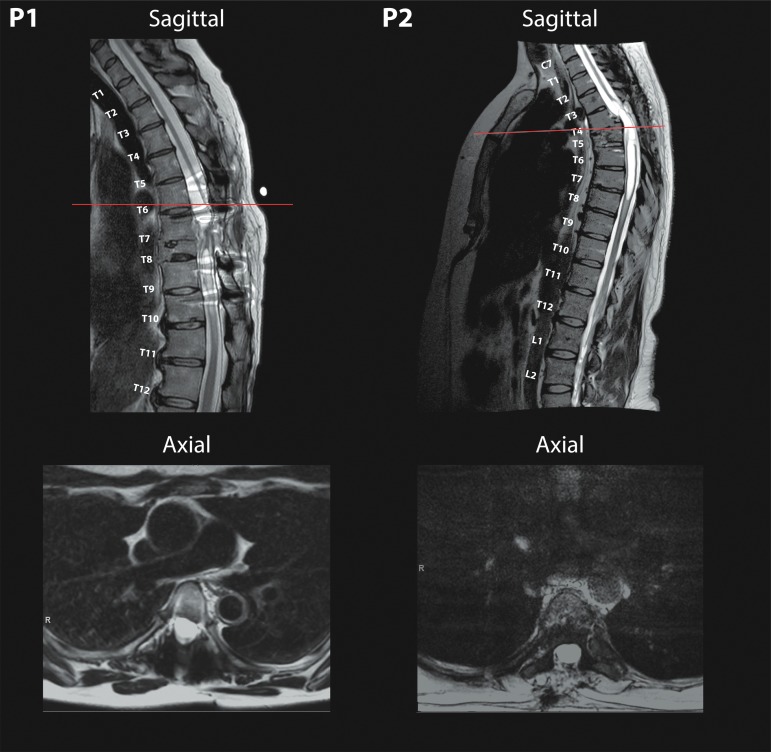
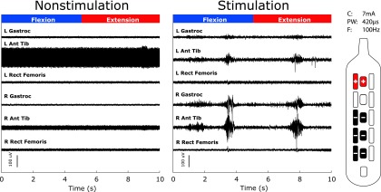
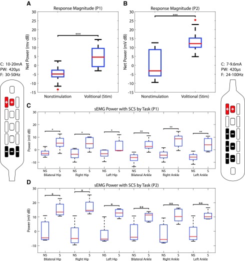
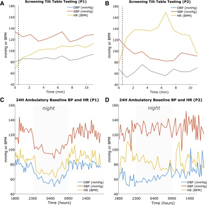
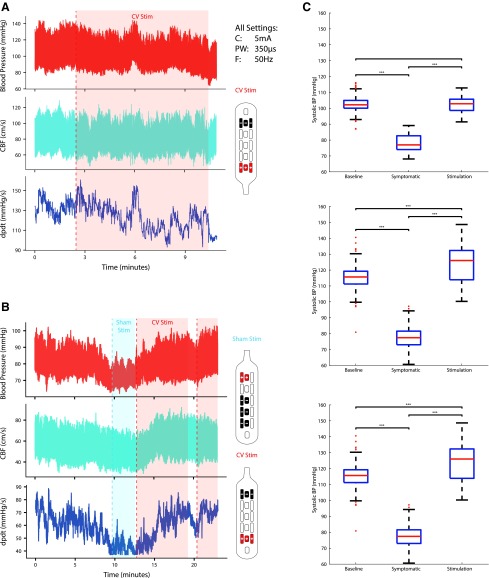
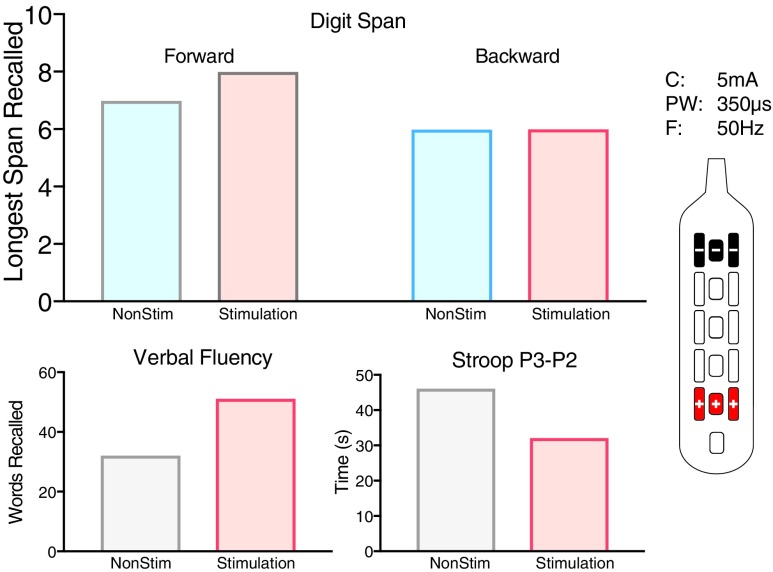
Epidural Spinal Cord Stimulation (eSCS) in combination with extensive rehabilitation has been reported to restore volitional movement in a select group of subjects after motor-complete spinal cord injury (SCI). Numerous questions about the generalizability of these findings to patients with longer term SCI have arisen, especially regarding the possibility of restoring autonomic function. To better understand the effect of eSCS on volitional movement and autonomic function, two female participants five and 10 years after injury at ages 48 and 52, respectively, with minimal spinal cord preservation on magnetic resonance imaging were implanted with an eSCS system at the vertebral T12 level. We demonstrated that eSCS can restore volitional movement immediately in two female participants in their fifth and sixth decade of life with motor and sensory-complete SCI, five and 10 years after sustaining severe radiographic injuries, and without prescribed or significant pre-habilitation. Both patients experienced significant improvements in surface electromyography power during a volitional control task with eSCS on. Cardiovascular function was also restored with eSCS in one participant with cardiovascular dysautonomia using specific eSCS settings during tilt challenge while not affecting function in a participant with normal cardiovascular function. Orgasm was achieved for the first time since injury in one participant with and immediately after eSCS. Bowel-bladder synergy improved in both participants while restoring volitional urination in one with eSCS. While numerous questions remain, the ability to restore some supraspinal control over motor function below the level of injury, cardiovascular function, sexual function, and bowel and bladder function should promote intense efforts to investigate and develop optimization strategies to maximize recovery in all participants with chronic SCI.

Keywords: autonomic; blood pressure; cardiovascular dysautonomia; spinal cord injury; spinal cord stimulation.


Conflict of interest statement

Dr. Darrow, Dr. Balser, Dr. Parr, and Dr. Samadani report a grant from MN State SCI/TBI Fund, grants from Abbott (Donation of Equipment) during the conduct of the study; Dr. Phillips and Dr. Krassioukov report grants from the Rick Hansen Institute during the conduct of the study. For Dr. Netoff, no competing financial interests exist.

Figures

<b>FIG. 1.</b>
FIG. 1.
Severe spinal cord injury with associated myelomalacia in both Participant 1 (left) and Participant 2 (right) from T8 and T4/5 injuries, respectively, in midsagittal planes of T2-weighted pre-operative magnetic resonance imaging. Axial slices (bottom) demonstrate minimal remaining spinal cord tissue with corresponding scout lines present on sagittal planes.
<b>FIG. 2.</b>
FIG. 2.
Surface electromyography (sEMG) during volitional tasks. Left panel shows raw baseline surface EMG during the bilateral ankle flexion and extension maneuver with spinal cord stimulation (SCS) off, and the right panel with SCS on. Appearance of large excursions in sEMG activity reflect voluntarily production of muscle contraction in response to verbal command (Participant 2). We observed recruitment of bilateral gastrocnemius and anterior tibialis without recruitment of the rectus femoris. The electrode configuration is shown to the right of the figure. Current (C) 7 mA, pulse-width (PW) 420 μs, and frequency (F) 100 Hz.
<b>FIG. 3.</b>
FIG. 3.
Differences in surface electromyography (sEMG) power. Pooled sEMG power between non-stimulation (NS) and stimulation (S) for the first five follow-up appointments for Participant 1 (P1) (A) and Participant 2 (P2) (B) demonstrating the power generated during volitional movement during Non-stimulation and stimulation (Volitional [stim]). The sEMG power for each maneuver during the first five follow-up appointments during NS and S (at intervals of approximately one month) for P1 (C) and P2 (D). Stimulator patterns for P1 are shown on the left, and patterns for P2 are shown on the right. In order of visit, P1 used frequencies (F) of 30, 30, 24, 50, and 30 Hz. Pulse-widths (PW) were 420 μs, and currents (C) were 13.5, 16.5, 10, 20, and 16 mA. P2 used frequencies of 30, 100, 30, 40, and 24 Hz. Pulse-width was 420 μs, and currents used were 7.8, 7, 7.5, 7.8, and 9.6 mA. Outliers (>2.7□) are denoted as red crosses. Asterisks denote statistical significance: *p < 0.05, **p < 0.01, ***p < 0.001.
<b>FIG. 4.</b>
FIG. 4.
Cardiovascular screening. Top: Tilt-table testing at screening for Participant 1 (P1) (A) and Participant 2 (P2) (B). The vertical dotted line indicates the time of tilt preceding the 10 min period. Notice the relative stability in P1 (A) compared with P2 (B). Bottom: Baseline ambulatory blood pressure and heart rate monitoring for 24 h for P1 (C) and P2 (D) with nocturnal hours denoted by gray. Notice the significant difference between P1 (C), who exhibits normal diurnal variation, and P2 (D), who exhibits little diurnal variation. DBP, diastolic blood pressure; SBP, systolic blood pressure; HR, heart rate.
<b>FIG. 5.</b>
FIG. 5.
Effect of spinal cord stimulation during tilt. (A) Tilt-table testing and stimulation had little effect on blood pressure (BP) for Participant 1 using stimulation for autonomic function (CV Stim). (B) Severe orthostatic hypotension during tilt-table evaluation in Participant 2 after 30 min of 70 degree tilt, acutely managed with application of CV Stim after failure of Sham Stim. Testing took place less than one month post-implantation. CBF (middle cerebral artery cerebral blood flow); dPdt (delta pressure/delta time—a measure of cardiac contractility). (C) Systolic BP changes. Systolic BP during supine rest (Baseline), symptomatic hypotension during tilt (Symptomatic), and steady state stimulation while tilted during three separate follow-up sessions for Participant 2. Stimulator patterns for Participants 1 and 2 are shown next to their respective graphs. Current (C) 5 mA, frequency (F) 50 Hz, pulse-width (PW) 350 μs were used for all electrode configurations.
<b>FIG. 6.</b>
FIG. 6.
Cognitive changes during tilt and stim. Cognitive performance during orthostatic challenge test in Participant 2 on Digit span (top), Verbal Fluency (bottom left), and Stroop (bottom right) between hypotensive episode and CV stimulation demonstrating improved cognitive function. Lower scores on the Stroop test are representative of increased cognitive function. Control, no stimulation. Stimulation, stimulator pattern shown on the right. Current (C) = 5 mA, frequency (F) 50 Hz, pulse-width (PW) = 350 μs were used for all electrode configurations.

References

    1. Shealy C.N., Mortimer J.T., and Reswick J.B. (1967). Electrical inhibition of pain by stimulation of the dorsal columns: preliminary clinical report. Anesth. Analg. 46, 489–491 - PubMed
    1. Shealy C.N., Mortimer J.T., and Hagfors N.R. (1970). Dorsal column electroanalgesia. J. Neurosurg. 32, 560–564 - PubMed
    1. Waltz J.M., Andreesen W.H., and Hunt D.P. (1987). Spinal cord stimulation and motor disorders. Pacing Clin. Electrophysiol. 10, 180–204 - PubMed
    1. Herman R., He J., D'Luzansky S., Willis W., and Dilli S. (2002). Spinal cord stimulation facilitates functional walking in a chronic, incomplete spinal cord injured. Spinal Cord 40, 65–68 - PubMed
    1. Harkema S., Gerasimenko Y., Hodes J., Burdick J., Angeli C., Chen Y., Ferreira C., Willhite A., Rejc E., Grossman R.G., and Edgerton V.R. (2011). Effect of epidural stimulation of the lumbosacral spinal cord on voluntary movement, standing, and assisted stepping after motor complete paraplegia: a case study. Lancet 377, 1938–1947 - PMC - PubMed

Publication types

MeSH terms