. 2019 Apr;291(1):196-202.
doi: 10.1148/radiol.2018180921. Epub 2019 Jan 22.

Automated Triaging of Adult Chest Radiographs with Deep Artificial Neural Networks

Affiliations

Automated Triaging of Adult Chest Radiographs with Deep Artificial Neural Networks

Mauro Annarumma et al. Radiology. 2019 Apr.

Erratum in

Abstract

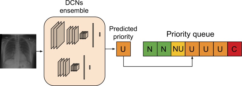
Purpose To develop and test an artificial intelligence (AI) system, based on deep convolutional neural networks (CNNs), for automated real-time triaging of adult chest radiographs on the basis of the urgency of imaging appearances. Materials and Methods An AI system was developed by using 470 388 fully anonymized institutional adult chest radiographs acquired from 2007 to 2017. The free-text radiology reports were preprocessed by using an in-house natural language processing (NLP) system modeling radiologic language. The NLP system analyzed the free-text report to prioritize each radiograph as critical, urgent, nonurgent, or normal. An AI system for computer vision using an ensemble of two deep CNNs was then trained by using labeled radiographs to predict the clinical priority from radiologic appearances only. The system's performance in radiograph prioritization was tested in a simulation by using an independent set of 15 887 radiographs. Prediction performance was assessed with the area under the receiver operating characteristic curve; sensitivity, specificity, positive predictive value (PPV), and negative predictive value (NPV) were also determined. Nonparametric testing of the improvement in time to final report was determined at a nominal significance level of 5%. Results Normal chest radiographs were detected by our AI system with a sensitivity of 71%, specificity of 95%, PPV of 73%, and NPV of 94%. The average reporting delay was reduced from 11.2 to 2.7 days for critical imaging findings (P < .001) and from 7.6 to 4.1 days for urgent imaging findings (P < .001) in the simulation compared with historical data. Conclusion Automated real-time triaging of adult chest radiographs with use of an artificial intelligence system is feasible, with clinically acceptable performance. © RSNA, 2019 Online supplemental material is available for this article. See also the editorial by Auffermann in this issue.


Figures

Figure 1:
Figure 1:
Flowchart shows different data sets used for training, learning, and testing. Approximately 8% of radiographs were critical, 40% urgent, 26% nonurgent, and 26% normal across the training, test, and validation data sets.
Figure 2:
Figure 2:
Example of radiologic report annotated by natural language processing system. “Entities” are highlighted with different colors, one for each semantic class. Arrows represent relationships between entities. Final annotation extracted by the rule-based system was “airspace opacification; pleural effusion/abnormality.”
Figure 3:
Figure 3:
Artificial intelligence prioritization system: When a chest radiograph is acquired, the deep learning architecture (consisting of two different deep convolutional neural networks [DCNs] operating at different input sizes) processes the image in real time and predicts its priority level (eg, urgent, in this example). Given the predicted priority, the image is then automatically inserted in a dynamic priority-based reporting queue. C = critical, N = normal, NU = nonurgent, U = urgent.
Figure 4:
Figure 4:
Receiver operating characteristic curve for normality prediction obtained by the artificial intelligence system. The system achieved an area under the receiver operating characteristic curve (AUC ) of 0.94.
Figure 5a:
Figure 5a:
Examples of correctly and incorrectly prioritized radiographs. (a) Radiograph was reported as showing large right pleural effusion (arrow). This was correctly prioritized as urgent. (b) Radiograph reported as showing “lucency at the left apex suspicious for pneumothorax.” This was prioritized as normal. On review by three independent radiologists, the radiograph was unanimously considered to be normal. (c) Radiograph reported as showing consolidation projected behind heart (arrow). The finding was missed by the artificial intelligence system, and the study was incorrectly prioritized as normal.
Figure 5b:
Figure 5b:
Examples of correctly and incorrectly prioritized radiographs. (a) Radiograph was reported as showing large right pleural effusion (arrow). This was correctly prioritized as urgent. (b) Radiograph reported as showing “lucency at the left apex suspicious for pneumothorax.” This was prioritized as normal. On review by three independent radiologists, the radiograph was unanimously considered to be normal. (c) Radiograph reported as showing consolidation projected behind heart (arrow). The finding was missed by the artificial intelligence system, and the study was incorrectly prioritized as normal.
Figure 5c:
Figure 5c:
Examples of correctly and incorrectly prioritized radiographs. (a) Radiograph was reported as showing large right pleural effusion (arrow). This was correctly prioritized as urgent. (b) Radiograph reported as showing “lucency at the left apex suspicious for pneumothorax.” This was prioritized as normal. On review by three independent radiologists, the radiograph was unanimously considered to be normal. (c) Radiograph reported as showing consolidation projected behind heart (arrow). The finding was missed by the artificial intelligence system, and the study was incorrectly prioritized as normal.
Figure 6:
Figure 6:
Mean reporting time from acquisition with artificial intelligence (AI) prioritization system compared with observed mean for critical radiographs. P values were obtained nonparametrically by using a null distribution (shown here), that is, a distribution of mean reporting time obtained under null hypothesis that order in which examinations are reported is not dependent on criticality class. The null distribution is generated by simulating 500 000 realizations of a randomized prioritization process, that is, the priority class in each realization is randomly assigned irrespective of image content.

Comment in

References

    1. Geras KJ, Wolfson S, Kim SG, Moy L, Cho K. High-resolution breast cancer screening with multi-view deep convolutional neural networks. Cornell University Library; https://arxiv.org/abs/1703.07047. Published June 28, 2018. Accessed August 15, 2018.
    1. Lakhani P, Sundaram B. Deep learning at chest radiography: automated classification of pulmonary tuberculosis by using convolutional neural networks. Radiology 2017;284(2):574–582. - PubMed
    1. Rumelhart DE, Hinton GE, Williams RJ. Learning representations by back- propagating errors. Nature 1986;323(6088):533–536.
    1. LeCun Y, Bengio Y, Hinton G. Deep learning. Nature 2015;521(7553):436–444. - PubMed
    1. Esteva A, Kuprel B, Novoa RA, et al. . Dermatologist-level classification of skin cancer with deep neural networks. Nature 2017;542(7639):115–118. - PMC - PubMed

Publication types

LinkOut - more resources