Review
. 2019 Jun;46 Suppl 21(Suppl Suppl 21):103-123.
doi: 10.1111/jcpe.13068.

Barrier membranes: More than the barrier effect?

Affiliations
Review

Barrier membranes: More than the barrier effect?

Omar Omar et al. J Clin Periodontol. 2019 Jun.

Abstract

Aim: To review the knowledge on the mechanisms controlling membrane-host interactions in guided bone regeneration (GBR) and investigate the possible role of GBR membranes as bioactive compartments in addition to their established role as barriers.

Materials and methods: A narrative review was utilized based on in vitro, in vivo and available clinical studies on the cellular and molecular mechanisms underlying GBR and the possible bioactive role of membranes.

Results: Emerging data demonstrate that the membrane contributes bioactively to the regeneration of underlying defects. The cellular and molecular activities in the membrane are intimately linked to the promoted bone regeneration in the underlying defect. Along with the native bioactivity of GBR membranes, incorporating growth factors and cells in membranes or with graft materials may augment the regenerative processes in underlying defects.

Conclusion: In parallel with its barrier function, the membrane plays an active role in hosting and modulating the molecular activities of the membrane-associated cells during GBR. The biological events in the membrane are linked to the bone regenerative and remodelling processes in the underlying defect. Furthermore, the bone-promoting environments in the two compartments can likely be boosted by strategies targeting both material aspects of the membrane and host tissue responses.


Conflict of interest statement

Omar Omar received grants from Osteology Foundation , grants from Hjalmar Svensson Foundation, during the conduct of the study. Ibrahim Elgali received a grant from Hjalmar Svensson Foundation, during the conduct of the study.Peter Thomsen received grants from Swedish Research Council , grants from ALF/LUA Research Grant , grants from IngaBritt and Arne Lundberg Foundation, grants from Vilhelm and Martina Lundgren Vetenskapsfond , grants from Area of Advance Materials of Chalmers and GU Biomaterials within the Strategic Research Area initiative launched by the Swedish Government, during the conduct of the study. Christer Dahlin received a grant from Osteology Foundation, during the conduct of the study.

Figures

Figure 1
Figure 1
Schematic diagram and clinical photographs of guided bone regeneration. (a) Simple schematic diagram showing the membrane and defect compartments in the guided bone regeneration procedure. (b–e) Serial clinical photographs of a horizontal bone defect treated by guided bone regeneration using a barrier membrane (titanium‐reinforced polytetrafluoroethylene (PTFE)): (b) the anterior maxilla with predominant bone loss is exposed, and a titanium tenting screw is inserted. (c) The titanium‐reinforced PTFE membrane is positioned and stabilized above the defect area. (d) After 7 months, the membrane is removed, and the previous defect has now been filled with bone. (e) Two titanium implants are inserted in the regenerated region and are subsequently connected with abutments and restored with final crowns (Courtesy of Drs Miranda‐Burgos & Dahlin)
Figure 2
Figure 2
Guided bone regeneration (GBR) using a synthetic, polytetrafluorethylene (PTFE) barrier membrane. The histological images (a and b) represent undecalcified, resin‐embedded and toluidine blue‐stained sections showing GBR using a titanium‐reinforced PTFE barrier membrane on a surgically created mandible defect in the dog model. (a) An orofacial section showing the pattern of bone formation under the membrane after 4 months of healing. The newly regenerated bone (NB) is formed in direct continuity with the host old bone (OB) under the barrier membrane, which separated the bone from the overlying oral mucosa (epithelium and connective tissue). (b) Under the periphery of the PTFE membrane, NB is formed on the porous surface of the PTFE. (c) The bar chart shows the amounts of radiopaque new bone within a rabbit 15‐mm cranial defect treated with a PTFE membrane and evaluated on radiographs at 1, 2, 3 and 5 weeks after surgery. The spatial analysis reveals the progressive increase in the amount of regenerated bone with respect to the amounts of total new bone (white bars), bone originating at the defect borders (hatched bars) and new bone formed as islands in the central region of the defect (black bars). The images (a and b) are adapted and republished with the permission of Quintessence Publishing Company Inc. from the Int J Oral Maxillofac Implants: Healing pattern of bone regeneration in membrane‐protected defects: a histological study in the canine mandible., Schenk RK, Buser D, Hardwick WR, Dahlin C., 9 (1), 1994; permission conveyed through the Copyright Clearance Center, Inc. The image in (c) is adapted and reprinted from the J Oral Maxillofac Surg, 53 (2), Hämmerle CH, Schmid J, Lang NP, Olah AJ., Temporal dynamics of healing in rabbit cranial defects using guided bone regeneration, 167‐74, copyright (1995), with permission from Elsevier [via the Copyright Clearance Center]
Figure 3
Figure 3
Cellular, molecular and structural events during guided bone regeneration (GBR) by collagen‐based membranes on surgically created bone defects in a rat model. (A) Immunohistochemical findings in decalcified and paraffin‐embedded sections showing GBR using a porcine type I/III collagen membrane consisting of a “compact” top part (*) and a “porous” bottom part (**) placed on a surgically created maxillary defect in the rat model after 2 weeks of healing. In (A), abundant newly formed bone (NB) is observed filling the defect, and an abundant cell infiltrate is observed in the porous part of the membrane facing the defect. The insert shows that the cells infiltrating the porous part of the porcine type I/III collagen membrane are positively stained for the bone proteins (a) alkaline phosphatase (ALP), (b) osteopontin (OPN) and (c) osteocalcin (OC), suggesting the active participation of the membrane‐associated cells in the bone regeneration process. (B and C) Histological images of undecalcified, resin‐embedded and toluidine blue‐stained sections showing that the application of a collagen membrane (derived from porcine small intestine extracellular matrix (ECM)) to a rat femur bone defect (B) results in structural restitution of the underlying defect with newly formed bone compared to the lesser restitution in the untreated defect (C). In the untreated sham defect (C), soft tissue invasion and poor restitution of the defect are evident. The histomorphometric analysis of the different regions of the defect (D and E) demonstrates a higher proportion of newly regenerated bone in the defect treated with the ECM collagen membrane than in the untreated sham defect, specifically in the top region of the defect directly underneath the membrane. The immunohistochemical analyses show that during GBR (exemplified here at 3 days), different cell types, including CD68‐positive macrophages (F) and periostin‐positive osteoprogenitor cells (G), are recruited and hosted within the ECM collagen membrane above the defect. Moreover, the immunohistochemical analysis reveals positive protein reactivity for the pro‐osteogenic, bone‐promoting growth factors FGF‐2 (H) and BMP‐2 (I) inside the membrane. The molecular analysis (qPCR) confirms the progressively increasing expression of the bone‐promoting growth factors FGF‐2 (J) and BMP‐2 (K) in conjunction with a temporal downregulation of the vascularization growth factor VEGF (L) in the membrane‐associated cells. The corresponding molecular qPCR analysis of the underlying defect reveals that the application of the ECM collagen membrane modulates the molecular activities of different healing processes, exemplified here by the pro‐inflammatory cytokine TNF‐α (M) and the bone formation gene OC (N), providing molecular evidence for membrane‐promoted bone healing and regeneration in the underlying defect. The significant correlations between the gene expression in the membrane and the gene expression in the underlying defect (insert table) demonstrate that the molecular activities in the two compartments are linked during the course of GBR. The upper panel of the figure (A and a, b c) is adapted and reprinted from Biomaterials, 26 (31), Taguchi Y, Amizuka N, Nakadate M, Ohnishi H, Fujii N, Oda K, Nomura S, Maeda T., A histological evaluation for guided bone regeneration induced by a collagenous membrane., 6158‐66, copyright (2005), with permission from Elsevier [via the Copyright Clearance Center]. The lower panel of the figure is adapted and reprinted from the Eur J Oral Sci, 125 (5), Elgali I, Omar O, Dahlin C, Thomsen P, Guided bone regeneration: materials and biological mechanisms revisited., 315–337, copyright (2017), published by John Wiley & Sons Ltd. under a Creative Commons license (CCBYNCND): https://creativecommons.org/licenses/by-nc-nd/4.0/
Figure 4
Figure 4
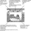
Schematic showing the membrane and bone defect compartments, both of which are amenable to potential strategies to enhance the clinical results of the GBR technique. The strategies include (1) the optimization of membrane material properties, (2) the incorporation of biological factors, natural elements and synthetic bioactive materials in the membrane, (3) the incorporation of antibiotic and antibacterial agents in the membrane, (4) the administration of osteoconductive and osteoinductive scaffolds/graft materials into the bone defect, and (5) the administration of biological cues into the bone defect. The figure is adapted and reprinted from Eur J Oral Sci, 125 (5), Elgali I, Omar O, Dahlin C, Thomsen P, Guided bone regeneration: materials and biological mechanisms revisited., 315–337, copyright (2017), published by John Wiley & Sons Ltd. under a Creative Commons licence (CCBYNCND): https://creativecommons.org/licenses/by-nc-nd/4.0/

References

    1. Aghaloo, T. L. , & Moy, P. K. (2007). Which hard tissue augmentation techniques are the most successful in furnishing bony support for implant placement? International Journal of Oral & Maxillofacial Implants, 22(Suppl), 49–70. - PubMed
    1. Ahn, Y. S. , Kim, S. G. , Kim, C. S. , Oh, J. S. , & Lim, S. C. (2012). Effect of guided bone regeneration with or without pericardium bioabsorbable membrane on bone formation. Oral Surgery, Oral Medicine, Oral Pathology and Oral Radiology, 114, S126–S131. 10.1016/j.oooo.2011.08.008 - DOI - PubMed
    1. Al‐Hezaimi, K. , Rudek, I. , Al‐Hamdan, K. S. , Javed, F. , Nooh, N. , & Wang, H. L. (2013). Efficacy of using a dual layer of membrane (dPTFE placed over collagen) for ridge preservation in fresh extraction sites: A micro‐computed tomographic study in dogs. Clinical Oral Implants Research, 24, 1152–1157. 10.1111/j.1600-0501.2012.02526.x - DOI - PubMed
    1. Al‐Kattan, R. , Retzepi, M. , Calciolari, E. , & Donos, N. (2017). Microarray gene expression during early healing of GBR‐treated calvarial critical size defects. Clinical Oral Implants Research, 28, 1248–1257. 10.1111/clr.12949 - DOI - PubMed
    1. Amar, S. , Chung, K. M. , Nam, S. H. , Karatzas, S. , Myokai, F. , & Van Dyke, T. E. (1997). Markers of bone and cementum formation accumulate in tissues regenerated in periodontal defects treated with expanded polytetrafluoroethylene membranes. Journal of Periodontal Research, 32, 148–158. 10.1111/j.1600-0765.1997.tb01397.x - DOI - PubMed

Publication types

Substances