

doi: 10.1109/MCG.2018.2877076.
CellPAINT: Interactive Illustration of Dynamic Mesoscale Cellular Environments
- PMID: 30668455
- PMCID: PMC6456043
- DOI: 10.1109/MCG.2018.2877076
Item in Clipboard
CellPAINT: Interactive Illustration of Dynamic Mesoscale Cellular Environments
IEEE Comput Graph Appl.
2018 Nov-Dec.
Abstract
CellPAINT allows nonexpert users to create interactive mesoscale illustrations that integrate a variety of biological data. Like popular digital painting software, scenes are created using a palette of molecular "brushes." The current release allows creation of animated scenes with an HIV virion, blood plasma, and a simplified T-cell.
Figures

Mesoscale scenes are often depicted using traditional computer graphics techniques (top), displaying global views of a section of the modeled environment and transitioning to an immersive view when zooming in (images created with cellVIEW, https://www.cg.tuwien.ac.at/cellview ). CellPAINT (bottom) depicts orthographic cross-sections, allowing the display of large mesoscale scenes and facile zooming to the level of individual molecules.
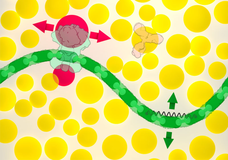
Soluble molecules, such as the Y-shaped antibody at upper right, are associated with circular colliders (shown in yellow). Membranes (green) and other articulated assemblies are created with modular components, with hinges between neighboring modules and springs to control large scale bending motion; the green arrows show the primary directions of motion available to a segment of membrane. Membrane-bound molecules, such as HIV envelope glycoprotein shown at upper left, are associated with two additional colliders (red) that only prevent overlap with the membrane, constraining the protein to move within the membrane (red arrows). A separate collider centered on the membrane protein (not shown) prevents overlap with soluble proteins.

The paths of insulin (the smallest protein in the cellPAINT plasma palette) and immunoglobulin M (one of the largest protein assemblies in the palette) are shown over 25 microseconds of random diffusive motion. The paths were traversed in 50 seconds of wall-clock time, so time was slowed by 2 million times.
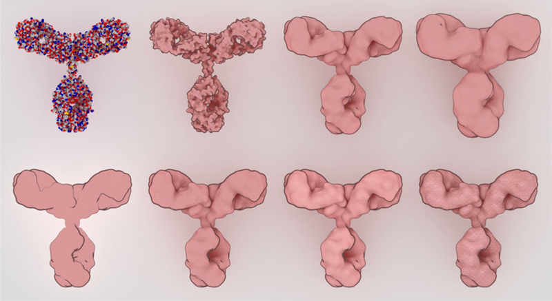
Sprites were designed with a simplified representation that still retains enough visual cues to highlight the shape and form of the molecule. At the top, a traditional spacefilling representation (left) is progressively smoothed with a blobby surface. At the bottom, non-photorealistic rendering parameters are explored, adding outlines, ambient occlusion, subtle specular highlights, and finally textures for the final sprite.
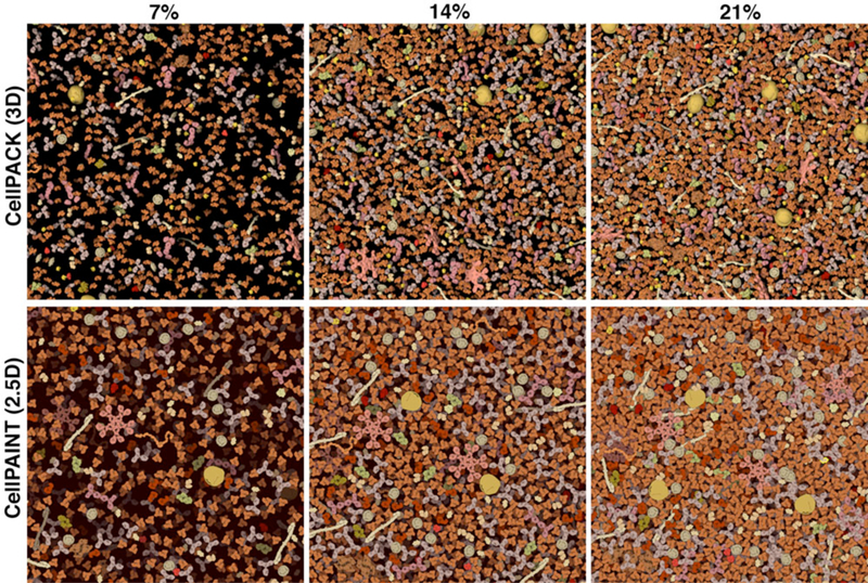
Visualization parameters were tuned by comparison with a 3D volume of blood plasma molecules. Images from 3D cellPACK models, with slab depth of 15 nanometers, are shown at the top and corresponding cellPAINT images are shown at the bottom. Three concentrations are shown: blood plasma has a typical concentration of about 7% (left), and the image at 21% (right) is more typical of cellular cytoplasm.
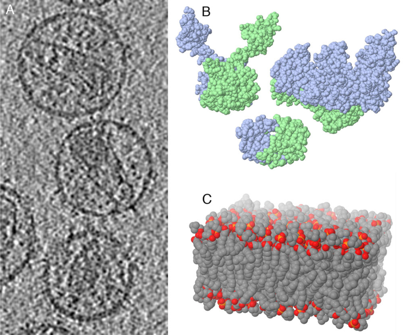
CellPAINT integrates experimental data from a variety of sources. Sources for illustrations of HIV include: a) an electron tomogram of mature virion; b) atomic structures of HIV proteins (integrase, reverse transcriptase and protease are shown here, PDB entries 1ex4, 1hys, 1hpv); c) computational simulations of lipid bilayers.
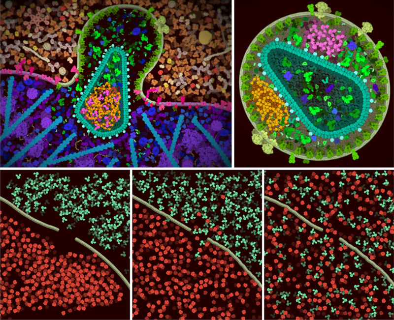
Sample screen captures from cellPAINT. (Top left) The distributed version of cellPAINT includes three palettes for creating a scene with HIV (green), blood plasma (tan and orange) and a simplified T-cell (blue and magenta). Here, we’ve created a scene after the virus has fused with the cell, releasing the capsid inside. The default colors have been changed to highlight integrase (magenta) and the viral genome with nucleocapsid (yellow and orange) inside the cone-shape capsid. A rendering option for vignetting is also used in the image. (Top right) Variations on this basic scene are also possible, such as a non-infectious virus that is formed when infected cells are treated with an integrase inhibitor. Notice that both the genome (orange) and integrase (magenta) are packaged outside the capsid in this aberrant form. (Bottom) CellPAINT may also be used to explore mesoscale properties, such as the diffusion of molecules through a semi-permeable membrane. Three time points are shown here: after roughly painting molecules in the scene, after 2 minutes (wall clock time) of diffusion at the room temperature, and after 2 hours of diffusion.

Cross sections of an entire mycoplasma cell integrate data from structural biology, microscopy and proteomics. An artistic watercolor rendering is shown at left and a 3D model generated by CellVIEW is shown at right..

User interface of cellPAINT..
References
-
- Berg HC, “Random Walks in Biology,” 1983, Princeton University Press, Princeton, New Jersey.
-
- Goodsell DS, “Illustrating the Machinery of Life: Viruses,” Biochem. Mol. Biol. Ed, vol. 40, no. 5, 2012, pp. 291–296. - PubMed
-
- Goodsell DS, “Illustrations of the HIV Life Cycle,” Curr. Top. Microbiol. Immunol, vol. 389, 2015, pp. 243–252. - PubMed
Publication types
MeSH terms
Grants and funding
LinkOut - more resources
Full Text Sources
Other Literature Sources
