. 2019 Jan 22;13(1):e0006948.
doi: 10.1371/journal.pntd.0006948. eCollection 2019 Jan.

A Biomphalaria glabrata peptide that stimulates significant behaviour modifications in aquatic free-living Schistosoma mansoni miracidia

Affiliations

A Biomphalaria glabrata peptide that stimulates significant behaviour modifications in aquatic free-living Schistosoma mansoni miracidia

Tianfang Wang et al. PLoS Negl Trop Dis. .

Abstract

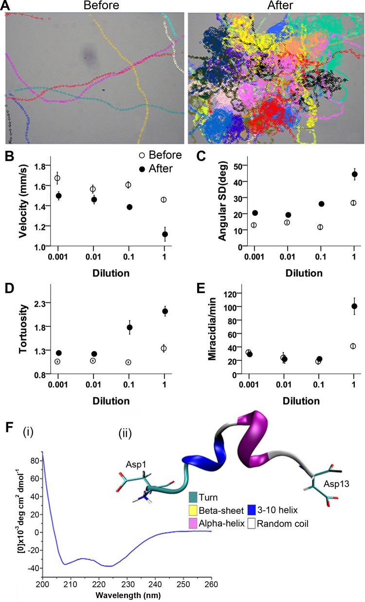
The human disease schistosomiasis (or bilharzia) is caused by the helminth blood fluke parasite Schistosoma mansoni, which requires an intermediate host, the freshwater gastropod snail Biomphalaria glabrata (the most common intermediate host). The free-swimming parasite miracidia utilise an excellent chemosensory sense to detect and locate an appropriate host. This study investigated the biomolecules released by the snail that stimulate changes in the behaviour of the aquatic S. mansoni miracidia. To achieve this, we have performed an integrated analysis of the snail-conditioned water, through chromatography and bioassay-guided behaviour observations, followed by mass spectrometry. A single fraction containing multiple putative peptides could stimulate extreme swimming behaviour modifications (e.g. velocity, angular variation) similar to those observed in response to crude snail mucus. One peptide (P12;-R-DITSGLDPEVADD-KR-) could replicate the stimulation of miracidia behaviour changes. P12 is derived from a larger precursor protein with a signal peptide and multiple dibasic cleavage sites, which is synthesised in various tissues of the snail, including the central nervous system and foot. P12 consists of an alpha helix secondary structure as indicated by circular dichroism spectroscopy. This information will be helpful for the development of approaches to manipulate this parasites life cycle, and opens up new avenues for exploring other parasitic diseases which have an aquatic phase using methods detailed in this investigation.


Conflict of interest statement

The authors have declared that no competing interests exist.

Figures

Fig 1
Fig 1. Workflow for identification of metabolites and peptides within B. glabrata conditioned water, then active biomolecule identification.
Snail conditioned water (SCW) was fractionated using HPLC followed by bioassay and identification using nanoLC tandem TripleTOF MS/MS.
Fig 2
Fig 2. Characterization of the behaviour of S. mansoni miracidia in response to raw mucus of B. glabrata.
(A) Arrangement of bioassay, including schematic illustration of video zone and corresponding area of observation. (B) Microscopic examination of site of application. Arrows show sites of miracidia for subsequent tracking. Asterisk indicates the pipette tip shadow. (C) Trajectories of miracidia in the presence of raw mucus. Different colours represent individual miracidium.
Fig 3
Fig 3. Identification of bioactive RP-HPLC fraction from B. glabrata conditioned water.
(A) Representative RP-HPLC chromatogram of SCW crude extracts of B. glabrata, with the active fraction highlighted. (B) Representative movement trajectories and density of miracidia 1 min before and 1 min after the addition of 2 μl of active fraction as a resuspended solution (protein concentration ~1 mg/ml). Each colour represents the swim-path of one tracked miracidium. See S5 Movie for the assay video. (C-F) Analyses of active fraction effect on miracidia behaviour measurements (means ± standard error). For each measurement type, paired t-tests (n = 6 for each) were used to compare measurements before and after addition of the bioactive HPLC fraction. Miracidia speed (C), angular standard deviation (D), track tortuosity (E) and rate of accumulation (F) before and after RP-HPLC fraction 20–25 min application.
Fig 4
Fig 4. Identification of B. glabrata P12 peptide and tissue expression.
(A) MS/MS spectrum of peptide P12—DITSGLDPEVADD-OH. (B) P12 precursor protein showing signal peptide (yellow), potential cleavage sites (red) and region of P12 (grey). (C) P12 gene expression in various B. glabrata tissues. TPM, transcripts per kilobase million mapped reads as determined by de novo RNA-seq [42] (CLC Genomic Workbench 9 software). APO, amebocyte-producing organ.
Fig 5
Fig 5. Bioassay and structure analysis of B. glabrata P12 peptide.
(A) Representative trajectories of miracidia movement before and after the addition of 2 μl of the P12 solution (0.742 μM) to the centre of the area. Each colour represents one indistinguishable miracidium individual. See S6 Movie for assay video. Miracidia speed (B), angular standard deviation (C), track tortuosity (D) and rate of accumulation (E) before and after P12 application at four different dilutions (means ± standard errors). (F) Structural analysis of P12. (i) Circular dichroism spectrum of P12, and (ii) representative structure of P12 at 306.4 K during the REMD simulation.

References

    1. Olveda DU, Li Y, Olveda RM, Lam AK, McManus DP, Chau TN, et al. Bilharzia in the Philippines: past, present, and future. International journal of infectious diseases: IJID: official publication of the International Society for Infectious Diseases. 2014;18:52–6. Epub 2013/11/12. 10.1016/j.ijid.2013.09.011 . - DOI - PubMed
    1. King CH. Parasites and poverty: the case of schistosomiasis. Acta tropica. 2010;113(2):95–104. Epub 2009/12/08. 10.1016/j.actatropica.2009.11.012 - DOI - PMC - PubMed
    1. Gray DJ, McManus DP, Li Y, Williams GM, Bergquist R, Ross AG. Schistosomiasis elimination: lessons from the past guide the future. The Lancet Infectious diseases. 2010;10(10):733–6. Epub 2010/08/14. 10.1016/S1473-3099(10)70099-2 . - DOI - PubMed
    1. WHO. 2018. Available from: http://www.who.int/schistosomiasis/epidemiology/table/en/.
    1. McManus DP, Dunne DW, Sacko M, Utzinger J, Vennervald BJ, Zhou XN. Schistosomiasis. Nature reviews Disease primers. 2018;4(1):13 Epub 2018/08/11. 10.1038/s41572-018-0013-8 . - DOI - PubMed

Publication types