. 2019 Mar 1;142(3):502-511.
doi: 10.1093/brain/awy339.

Blood-brain barrier pericytes as a target for HIV-1 infection

Affiliations

Blood-brain barrier pericytes as a target for HIV-1 infection

Luc Bertrand et al. Brain. .

Abstract

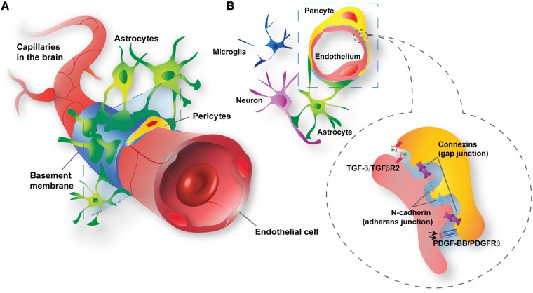
Pericytes are multifunctional cells wrapped around endothelial cells via cytoplasmic processes that extend along the abluminal surface of the endothelium. The interactions between endothelial cells and pericytes of the blood-brain barrier are necessary for proper formation, development, stabilization, and maintenance of the blood-brain barrier. Blood-brain barrier pericytes regulate paracellular flow between cells, transendothelial fluid transport, maintain optimal chemical composition of the surrounding microenvironment, and protect endothelial cells from potential harmful substances. Thus, dysfunction or loss of blood-brain barrier pericytes is an important factor in the pathogenesis of several diseases that are associated with microvascular instability. Importantly, recent research indicates that blood-brain barrier pericytes can be a target of HIV-1 infection able to support productive HIV-1 replication. In addition, blood-brain barrier pericytes are prone to establish a latent infection, which can be reactivated by a mixture of histone deacetylase inhibitors in combination with TNF. HIV-1 infection of blood-brain barrier pericytes has been confirmed in a mouse model of HIV-1 infection and in human post-mortem samples of HIV-1-infected brains. Overall, recent evidence indicates that blood-brain barrier pericytes can be a previously unrecognized HIV-1 target and reservoir in the brain.

Keywords: HIV; HIV reservoirs; HIV-associated neurocognitive disorder; blood–brain barrier pericytes; neuroinfection.


Figures

Figure 1
Figure 1
Structural and molecular blood–brain barrier pericyte connections within the neurovascular unit. (A) Blood–brain barrier pericytes (yellow) and endothelial cells (red) share the basement membrane (sky blue) or are in direct contacts. In addition, blood–brain barrier pericytes are surrounded by glial cells (astrocytes, green; microglia, dark blue) and neurons (purple). (B) Blood–brain barrier pericytes and endothelial cells communicate with each other by direct contact (gap and adherens junctions) or through signalling pathways, such as platelet-derived growth factor BB (PDGF-BB)/PDGFRβ and transforming growth factor-β (TGF-β)/type 2 TGF-β receptor (TGFβR2).
Figure 2
Figure 2
Blood–brain barrier pericyte markers. Standardized characterization of non-infected (A) and HIV-1-infected (B) pericytes. Infections were performed by incubating pericytes with HIV-1 (NL4-3 strain) in the amount of 60 ng p24/ml for 48 h. Top: Phase contrast imaging; middle three panels represent immunofluorescence staining for the three distinct pericyte markers; namely, PDGFRβ, NG2, and αSMA (all in green). Bottom: Merged images of triple stained pericytes for these markers (PDGFRβ, green; NG2, blue; and αSMA, red). Nuclei were stained with Hoechst (blue). Scale bars = 200 µm (brightfield) and 50 µm (PDGFRβ, NG2, and αSMA).
Figure 3
Figure 3
HIV-1 replication and reactivation in human primary blood–brain barrier pericytes. (A) Quantification of p24 release into culture media at the indicated days post-infection with different HIV-1 strains, namely NL4-3, YU-2, or 49.5, in the amount of 60 ng p24/ml. The tropism (X4 or R5) of these strains is indicated on the graph. Day 0 indicates the start of infection. Data represent mean ± standard error of the mean (SEM) from three independent experiments, (3 × 10⁵ pericytes per replicate, total n = 9 per group). No p24 levels were detected in the non-infected (NI) group. (B) Representative images of p24 immunoreactivity at Day 2 post-infection with HIV-1 NL4-3 (60 ng p24/ml; orthogonal view in the merged image of HIV-1 NL4-3 group). No p24 levels were detected in NI group. Nuclei (blue, Hoechst staining), p24 (green, HIV-1 marker) and membranes (red, DiI staining). Scale bar = 10 µm. (C) Blood–brain barrier pericytes were infected with HIV-1 NL4-3 as in Fig. 3A (3 × 10⁵, 60 ng p24/ml), washed and incubated for 7 days. HIV-1 p24 release from HIV-1-infected pericytes and HIV-1 DNA integration into their genome as quantified by droplet digital PCR (ddPCR). Note that a decrease in active production of p24 is associated with elevated integration of the HIV-1 genome into the host genome. *P < 0.05 versus Day 3 post-infection. (D and E) On Day 8 post-infection, 3 × 10⁵ pericytes were exposed to the indicated factors for 3 days and then assayed for either (D) HIV-1 p24 by ELISA or (E) HIV-1 RNA using RT-qPCR. Results are reflected by minimum and maximum box and whisker plots. The HIV-1 reactivation factors were used at the following concentrations: TNF, 100 U/ml; SAHA, 10 µM; apicidin, 1 µg/ml. *P < 0.05 versus HIV-1; **P < 0.01 versus HIV-1; ***P < 0.001 versus HIV-1. AC were adapted from Cho et al. (2017).
Figure 4
Figure 4
Infection of blood–brain barrier pericytes by HIV in vivo. (A) Male C57BL/6 mice were infected with EcoHIV-1/NDK (1 µg of p24) via infusion through the internal carotid artery, while control animals (non-infected, NI) received saline. Mice were sacrificed at Day 7 post-infection. Brain microvessels were isolated and immunostained for PDGFRβ (green, arrow, marker of pericytes) and HIV-1 protein p24 (red, arrow, marker of active HIV-1 infection). Scale bars = 20 µm. (B) Triple staining of infected microvessels from (A) for PDGFRβ (green, arrow), p24 (red, arrow), and CD31 (blue, marker for microvessel endothelial cells). Arrows denote area of co-localization solely between p24 and PDGFRβ but not with CD31, indicating that blood–brain barrier pericytes but not brain endothelial cells, are infected. (C) In situ RT-PCR assay using fluorescently-labelled primers against spliced HIV-1 Rev mRNA (blue, arrow), HIV-1 gag (NDK, red, arrow), and NG2 spliced mRNA (green, pericyte marker). Focal signal indicates area of cDNA and DNA amplification. No signal for spliced Rev and NDK were observed in non-infected mice. (D) Brain samples (frontal cortex; 0.5 cm3 each) from three healthy (non-infected, NI) and three HIV-1-infected patients with HIV encephalopathy were processed to isolate microvessels. Microvessels from each brain sample were spread on around 30 slides, each slide containing ~100–150 microvessels of varying sizes and number of associated pericytes. Samples were then immunostained for PDGFRβ (green, marker of pericytes) and HIV-1 protein p24 (red, marker of active HIV-1 infection). Approximately five infected pericytes were clearly detected on each slide containing microvessels from infected brains. Arrows indicate area of co-localization of p24 and PDGFRβ. Scale bars = 10 µm. Arrow indicates area of co-localization.

References

    1. Armulik A, Genové G, Betsholtz C.. Pericytes: developmental, physiological, and pathological perspectives, problems, and promises. Dev Cell 2011; 21: 193–215. - PubMed
    1. Armulik A, Genové G, Mäe M, Nisancioglu MH, Wallgard E, Niaudet C, et al.Pericytes regulate the blood-brain barrier. Nature 2010; 468: 557–61. - PubMed
    1. Arnold TD, Niaudet C, Pang MF, Siegenthaler J, Gaengel K, Jung B, et al.Excessive vascular sprouting underlies cerebral hemorrhage in mice lacking αVβ8-TGFβ signaling in the brain. Development 2014; 141: 4489–99. - PMC - PubMed
    1. Asahina K, Zhou B, Pu WT, Tsukamoto H.. Septum transversum-derived mesothelium gives rise to hepatic stellate cells and perivascular mesenchymal cells in developing mouse liver. Hepatology 2011; 53: 983–95. - PMC - PubMed
    1. Attwell D, Buchan AM, Charpak S, Lauritzen M, Macvicar BA, Newman EA. Glial and neuronal control of brain blood flow. Nature 2010; 468: 232–43. - PMC - PubMed

Publication types