. 2019 Apr 1;29(4):1778-1788.
doi: 10.1093/cercor/bhy348.

Reduced White Matter Fiber Density in Autism Spectrum Disorder

Affiliations

Reduced White Matter Fiber Density in Autism Spectrum Disorder

Dennis Dimond et al. Cereb Cortex. .

Abstract

Differences in brain networks and underlying white matter abnormalities have been suggested to underlie symptoms of autism spectrum disorder (ASD). However, robustly characterizing microstructural white matter differences has been challenging. In the present study, we applied an analytic technique that calculates structural metrics specific to differently-oriented fiber bundles within a voxel, termed "fixels". Fixel-based analyses were used to compare diffusion-weighted magnetic resonance imaging data from 25 individuals with ASD (mean age = 16.8 years) and 27 typically developing age-matched controls (mean age = 16.9 years). Group comparisons of fiber density (FD) and bundle morphology were run on a fixel-wise, tract-wise, and global white matter (GWM) basis. We found that individuals with ASD had reduced FD, suggestive of decreased axonal count, in several major white matter tracts, including the corpus callosum (CC), bilateral inferior frontal-occipital fasciculus, right arcuate fasciculus, and right uncinate fasciculus, as well as a GWM reduction. Secondary analyses assessed associations with social impairment in participants with ASD, and showed that lower FD in the splenium of the CC was associated with greater social impairment. Our findings suggest that reduced FD could be the primary microstructural white matter abnormality in ASD.

Keywords: autism spectrum disorder; fiber density; fixel-based analysis; social impairment; white matter.


Figures

Figure 1.
Figure 1.
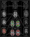
Methods schematic. Visual overview of the processing steps involved in the fixel- and voxel-based analyses utilized in the study. Processing steps specific to the fixel-based analysis are shown on the left panel, while those specific to the voxel-based analysis are shown on the right panel. The center panel depicts steps involved in both analyses. DWIs = diffusion-weighted images; FOD = fiber orientation distribution; FA = fractional anisotropy; MD = mean diffusivity; FD = fiber density; FC = fiber cross-section; FDC = fiber density and cross-section; FBA = fixel-based analysis; VBA = voxel-based analysis.
Figure 2.
Figure 2.
Analysis masks and tracts. (A) Voxel analysis mask visualized as a red overlay on 2D slices of the study template FOD image. (B) Fixel analysis mask overlaid onto a 2D sagittal slice of the study template FOD image (center), with fixels depicted as black lines. Windows to the left and right show zoomed in images of the regions enclosed in white squares within the central window. (C) Tracts that were utilized in defining the analysis masks (cingulum, AF, SLF, ILF, IFOF, UF, and corpus callosum), visualized as 3D streamlines within 2D slices of the study template FOD image. Streamlines are colored by direction: red = left–right; green = anterior–posterior; blue = superior–inferior.
Figure 3.
Figure 3.
Reduced fixel-wise fiber density in participants with ASD compared with typical controls. Fixels in which fiber density was reduced in participants with ASD relative to controls, visualized as 3D fixels overlaid onto a 3D brain image of the study template FOD. (A) Fixels are colored by direction: red = left–right; green = anterior–posterior; blue = superior–inferior. (B) Fixels are colored by percent reduction in fiber density relative to the typically developing (TD) controls.
Figure 4.
Figure 4.
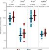
Reduced tract mean fiber density in participants with ASD compared with typical controls. Individual participant mean FD values for tracts in which average FD was reduced in individuals with ASD relative to controls. ∗ indicates tracts that were significantly different after FDR correction (q < 0.05). CC = corpus callosum; R-AF = right arcuate fasciculus; R-UF = right uncinate fasciculus; GWM = global white matter.
Figure 5.
Figure 5.
Negative correlation between fixel-wise fiber density and social impairment in participants with ASD. Fixels in which fiber density correlated negatively with SRS-2 scores in participants with ASD, visualized as 3D fixels overlaid onto a 2D slice of the study template FOD image. Fixels are colored by direction: red = left–right; green = anterior–posterior; blue = superior–inferior.

References

    1. Alexander AL, Lee JE, Lazar M, Boudos R, DuBray MB, Oakes TR, Miller JN, Lu J, Jeong EK, McMahon WM, et al. . 2007. Diffusion tensor imaging of the corpus callosum in Autism. Neuroimage. 34:61–73. - PubMed
    1. Ameis SH, Catani M. 2015. Altered white matter connectivity as a neural substrate for social impairment in Autism Spectrum Disorder. Cortex. 62:158–181. - PubMed
    1. American Psychiatric Association 2013. Diagnostic and Statistical Manual of Mental Disorders. Washington, DC: American Psychiatric Association.
    1. Andersson JLR, Graham MS, Drobnjak I, Zhang H, Filippini N, Bastiani M. 2017. Towards a comprehensive framework for movement and distortion correction of diffusion MR images: Within volume movement. Neuroimage. 152:450–466. - PMC - PubMed
    1. Andersson JL, Graham MS, Zsoldos E, Sotiropoulos SN. 2016. Incorporating outlier detection and replacement into a non-parametric framework for movement and distortion correction of diffusion MR images. Neuroimage. 141:556–572. - PubMed

Publication types

Grants and funding