. 2019 Apr;33(4):5599-5614.
doi: 10.1096/fj.201802063RR. Epub 2019 Jan 22.

MicroRNA-135a-3p regulates angiogenesis and tissue repair by targeting p38 signaling in endothelial cells

Affiliations

MicroRNA-135a-3p regulates angiogenesis and tissue repair by targeting p38 signaling in endothelial cells

Basak Icli et al. FASEB J. 2019 Apr.

Abstract

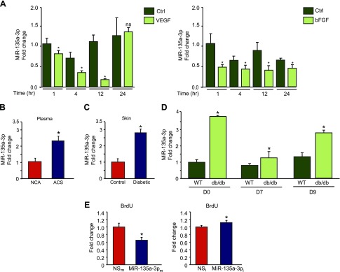
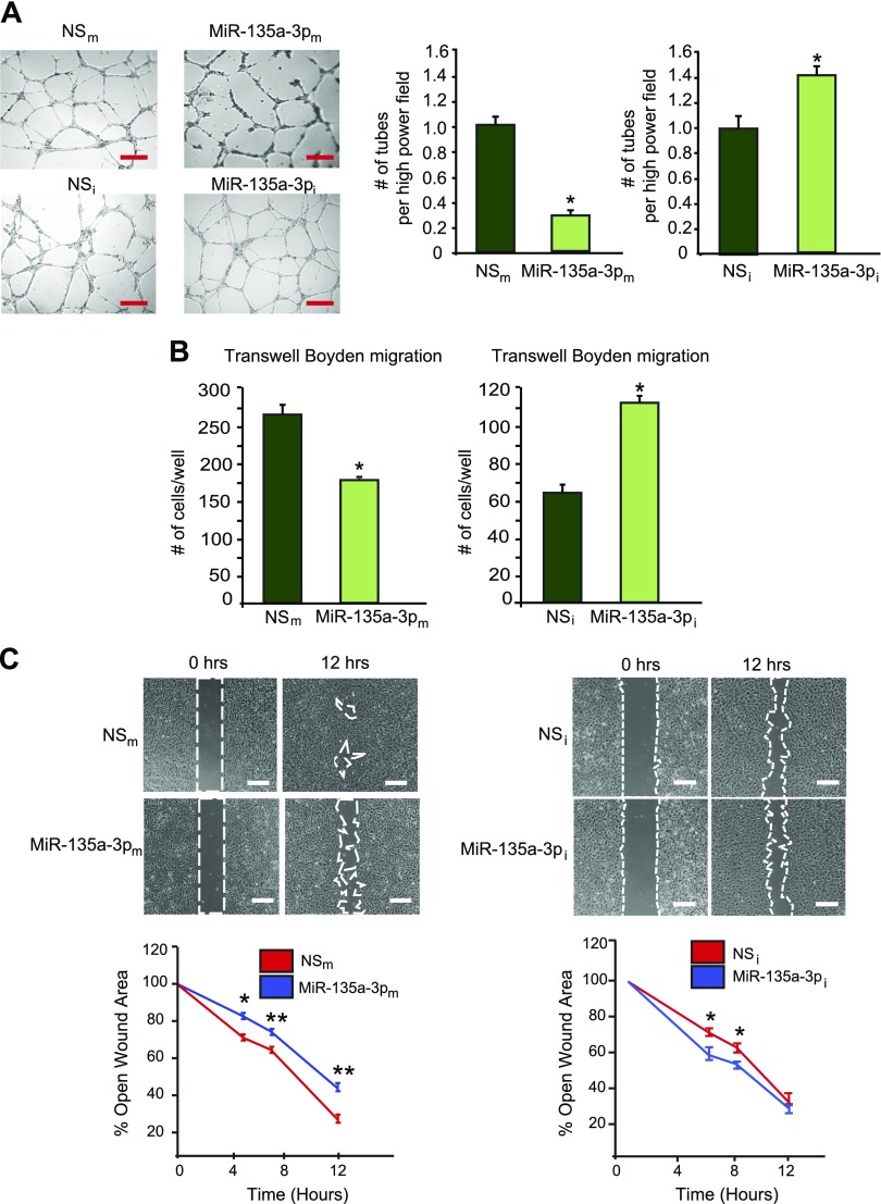
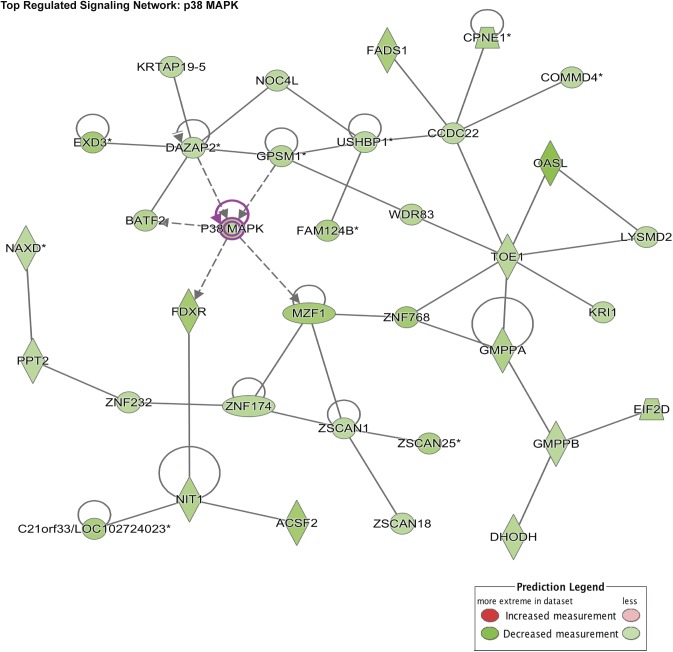
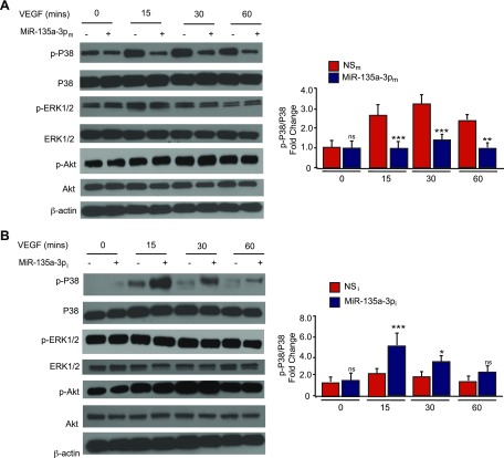
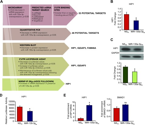
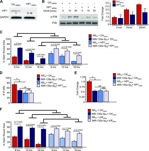
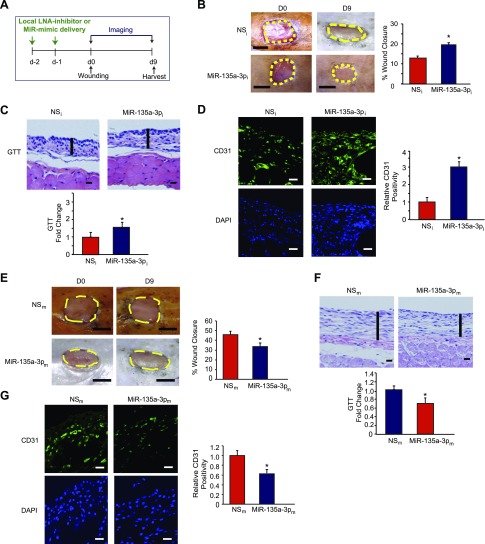
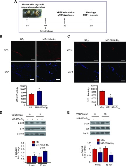
Angiogenesis is a critical process in repair of tissue injury that is regulated by a delicate balance between pro- and antiangiogenic factors. In disease states associated with impaired angiogenesis, we identified that miR-135a-3p is rapidly induced and serves as an antiangiogenic microRNA (miRNA) by targeting endothelial cell (EC) p38 signaling in vitro and in vivo. MiR-135a-3p overexpression significantly inhibited EC proliferation, migration, and network tube formation in matrigel, whereas miR-135-3p neutralization had the opposite effects. Mechanistic studies using transcriptomic profiling, bioinformatics, 3'-UTR reporter and miRNA ribonucleoprotein complex -immunoprecipitation assays, and small interfering RNA dependency studies revealed that miR-135a-3p inhibits the p38 signaling pathway in ECs by targeting huntingtin-interacting protein 1 (HIP1). Local delivery of miR-135a-3p inhibitors to wounds of diabetic db/db mice markedly increased angiogenesis, granulation tissue thickness, and wound closure rates, whereas local delivery of miR-135a-3p mimics impaired these effects. Finally, through gain- and loss-of-function studies in human skin organoids as a model of tissue injury, we demonstrated that miR-135a-3p potently modulated p38 signaling and angiogenesis in response to VEGF stimulation by targeting HIP1. These findings establish miR-135a-3p as a pivotal regulator of pathophysiological angiogenesis and tissue repair by targeting a VEGF-HIP1-p38K signaling axis, providing new targets for angiogenic therapy to promote tissue repair.-Icli, B., Wu, W., Ozdemir, D., Li, H., Haemmig, S., Liu, X., Giatsidis, G., Cheng, H. S., Avci, S. N., Kurt, M., Lee, N., Guimaraes, R. B., Manica, A., Marchini, J. F., Rynning, S. E., Risnes, I., Hollan, I., Croce, K., Orgill, D. P., Feinberg, M. W. MicroRNA-135a-3p regulates angiogenesis and tissue repair by targeting p38 signaling in endothelial cells.

Keywords: VEGF; diabetic wounds; human organoid.


Conflict of interest statement

The authors thank Lay-Hong Ang (Beth Israel Deaconess Medical Center, Boston, MA, USA) for confocal microscopy technical assistance, and the Harvard Digestive Disease Center and U.S. National Institutes of Health (NIH) National Institute of Diabetes and Digestive and Kidney Diseases Grant P30DK034854 This work was supported by the NIH National Heart, Lung, and Blood Institute Grants HL115141, HL117994, and HL134849, and NIH National Institute of General Medical Sciences Grant GM115605 (to M.W.F.); the Arthur K. Watson Charitable Trust (to M.W.F.); the Dr. Ralph and Marian Falk Medical Research Trust (Bank of America, N.A., Trustee; to M.W.F.); the American Heart Association Grant 18SFRN33900144 (to M.W.F.); the American Diabetes Association Grant 1-16-JDF-046 (to B.I.); the Watkins Discovery Award (to B.I.); the Lerner Young Investigator Award (to B.I.); a Tübitak Predoctoral Scholarship (to D.O.); and a grant from South-Eastern Regional Health Authorities and from Norwegian Women’s Public Health Association, Norway (to I.H.). The authors declare no conflicts of interest.

Figures

Figure 1
Figure 1
MiR-135a-3p is regulated by proangiogenic stimuli and inhibits EC growth. A) Real-time qPCR analysis of miR-135-3p expression in response to VEGF (25 ng/ml) and bFGF (25 ng/ml) in HUVECs. *P < 0.01 compared to control (Ctrl). B) Circulating miR-135a-3p levels are increased in plasma from human subjects with ACS (n = 20) compared to subjects with normal coronary angiograms (NCA) (n = 40). *P < 0.05 compared to normal coronary angiogram. C) MiR-135a-3p expression is increased in skin of patients with diabetes (n = 13) compared to nondiabetic controls (n = 10). *P < 0.05 compared to controls. D) Wild-type (WT) and db/db mice underwent punch-biopsy wounding of the dorsal skin, and wounds were collected for qPCR analyses for miR-135a-3p on the indicated days after wounding. E) HUVECs transfected with NSm, miR-135a-3pm, miR inhibitor negative control (NSi), or miR-135a-3p inhibitor (miR-135a-3pi) were subjected to BrdU cell proliferation assay. Ns, nonsignificant. All data represent means ± sem. *P < 0.05 compared to controls.
Figure 2
Figure 2
MiR-135a-3p inhibits proangiogenic functions in ECs in vitro. HUVECs transfected with NSm, miR-135a-3pm, miR inhibitor negative control (NSi), or miR-135a-3p inhibitor (miR-135a-3pi) were subjected to tube-like network formation in matrigel (A); EC migration in transwell Boyden chambers (B); scratch assay (C); n = 6/group. All data represent means ± sem. Scale bars: 150 µm (A); 100 µm (C). *P < 0.05 compared to NS, **P < 0.001 compared to NS.
Figure 3
Figure 3
Bioinformatics and miR-135a-3p gene profiling predicts p38 as a targeted signaling pathway. Gene ontology and GSEA predicted p38 MAPK signaling pathway to be the top regulated signaling network regulated by miR-135a-3p.
Figure 4
Figure 4
MiR-135a-3p regulates the expression of downstream P38 signaling in ECs. HUVECs transfected with NSm or miR-135a-3pm (A) or miR inhibitor negative control (NSi) or miR-135a-3p inhibitor (miR-135a-3pi) (B), and stimulated with VEGF (50 ng/ml) at the given time points were subjected to Western analysis using antibodies to p-p38, p38, p-ERK1/2, ERK1/2, p-Akt, Akt, and β-actin (n = 3–5 experiments). Ns, nonsignificant. All data represent means ± sem. Differences among groups were analyzed by using 1-way ANOVA. *P < 0.05, **P < 0.005, ***P < 0.001 compared to controls.
Figure 5
Figure 5
HIP1 is a bona fide target of miR-135a-3p in ECs. A) Discovery and validation of miR-135a-3p target genes. HUVECs transfected with NSm and miR-135a-3pm were subjected to microarray gene profiling. Potential gene targets were further narrowed down by sequential use of bioinformatics and prediction algorithms, real-time qPCR, Western blot analyses, 3′-UTR reporter studies, and miRNP-IP analysis. B, C) HUVECs transfected with NSm or miR-135a-3pm were subjected to real-time qPCR for HIP1 expression (B) or Western blot analyses using antibodies to HIP1 and GAPDH (n = 3 experiments) (C). D) Luciferase activity of HIP1 3′-UTR normalized to total protein was quantified in HUVECs transfected with NSm or miR-135a-3pm (n = 3 experiments). E) MiRNP-IP analysis of enrichment of HIP1 mRNA in HUVECs transfected with NSm or miR-135a-3pm. RT-qPCR was performed to detect HIP1 or SMAD1. *P < 0.01. ns, nonsignificant. Results are representative of n = 3 replicates/group and 2 independent experiments. All data represent means ± sem.
Figure 6
Figure 6
SiRNA-mediated knockdown of HIP1 recapitulates miR-135-3p functional effects in ECs. A, B) HUVECs were transfected with siRNA to HIP1 or scrambled control (Ctrl) siRNA. Protein expression was determined by Western analysis under baseline conditions (A) or in response to VEGF (50 ng/ml) treatment (B) using antibodies to HIP1, p-P38, P38, and GAPDH (n = 2 experiments). *P < 0.05, **P < 0.001. CE) HUVECs were transfected with siRNA to HIP1 or scrambled Ctrl siRNA in the presence of NSm or miR-135a-3pm. EC functional angiogenic assays were performed for scratch assay (C), Boyden transwell migration (D), or proliferation by BrdU assay (E). *P < 0.01. F) HUVECs were transfected with siRNA to p38K or siRNA control in the presence of NSm or miR-135a-3pm and EC scratch assays were performed. Ns, nonsignificant. Results are representative of n = 3 replicates/group. All data represent means ± sem. Differences among groups were analyzed by using 1-way ANOVA. *P < 0.01.
Figure 7
Figure 7
Local delivery of LNA-anti-miR-135a-3p promotes wound healing in db/db mice. A) After 2 local injections in mice of LNA-anti-miR-135a-3p (MiR-135a-3pi) or scrambled NS control LNA-anti-miRs (NSi) (n = 11–12/group), mice underwent dorsal skin wounding. BD) Wound analyses included: wound closure areas (B), GTT (C), and confocal immunofluorescence staining for CD31 (D). EG) After 2 local injections in mice of MiR-135a-3 PM or scrambled NS control miRs (NSm) (n = 10/group), mice underwent dorsal skin wounding. Wound analyses included: wound closure areas (E), GTT (F), and confocal immunofluorescence staining (G) for CD31. All data represent means ± sem. Scale bars: 5 mm (B, E); 500 μm (C, F); 50 μm (D, G). *P < 0.05.
Figure 8
Figure 8
Inhibition of miR-135a-3p promotes angiogenesis in human skin organoids. A) Punch biopsies of human skin were embedded into a collagen matrix, transfected with miR inhibitor negative control (NSi), miR-135a-3p inhibitor (miR-135a-5pi), NSm, or miR-135a-3pm, and cultured for indicated number of days. B, C) Human skin organoids were transduced with the indicated miRNAs and cultured for 9 d followed by confocal immunofluorescence staining for CD31. D, E) Human skin organoids (n = 3–6) were transduced with the indicated miRNAs and cultured for 3 d. Human skin organoids were treated with VEGF (50 ng/ml) followed by Western blot analyses for p-p38, p38, and β-actin at the indicated times. All data represent means ± sem. Scale bars: 50 μm (B); 25 μm (C). *P < 0.05 (1-way ANOVA).

References

    1. Krishna S. M., Moxon J. V., Golledge J. (2015) A review of the pathophysiology and potential biomarkers for peripheral artery disease. Int. J. Mol. Sci. 16, 11294–11322 - PMC - PubMed
    1. Falanga V. (2005) Wound healing and its impairment in the diabetic foot. Lancet 366, 1736–1743 - PubMed
    1. Prabhu S. D., Frangogiannis N. G. (2016) The biological basis for cardiac repair after myocardial infarction: from inflammation to fibrosis. Circ. Res. 119, 91–112 - PMC - PubMed
    1. Potente M., Gerhardt H., Carmeliet P. (2011) Basic and therapeutic aspects of angiogenesis. Cell 146, 873–887 - PubMed
    1. Kusumanto Y. H., van Weel V., Mulder N. H., Smit A. J., van den Dungen J. J., Hooymans J. M., Sluiter W. J., Tio R. A., Quax P. H., Gans R. O., Dullaart R. P., Hospers G. A. (2006) Treatment with intramuscular vascular endothelial growth factor gene compared with placebo for patients with diabetes mellitus and critical limb ischemia: a double-blind randomized trial. Hum. Gene Ther. 17, 683–691 - PubMed

Publication types

MeSH terms

Substances