. 2019 Mar:197:244-254.
doi: 10.1016/j.biomaterials.2019.01.020. Epub 2019 Jan 11.

A new class of biological materials: Cell membrane-derived hydrogel scaffolds

Affiliations

A new class of biological materials: Cell membrane-derived hydrogel scaffolds

Zhiyuan Fan et al. Biomaterials. 2019 Mar.

Abstract

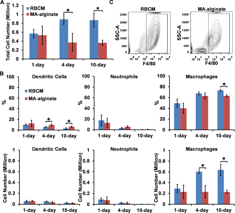
Biological materials are superior to synthetic biomaterials in biocompatibility and active interactions with cells. Here, a new class of biological materials, cell membrane-derived hydrogel scaffolds are reported for harnessing these advantages. To form macroporous scaffolds, vesicles derived from red blood cell membranes (RBCMs) are chemically crosslinked via cryogelation. The RBCM scaffolds with a pore size of around 70 μm are soft and injectable. Highly biocompatible scaffolds are typically made of superhydrophilic polymers and lack the ability to encapsulate and release hydrophobic drugs in a controlled manner. However, hydrophobic molecules can be efficiently encapsulated inside RBCM scaffolds and be sustainedly released. RBCM scaffolds show low neutrophil infiltration after subcutaneous injection in mice, and a significantly higher number of infiltrated macrophages than methacrylate alginate (MA-alginate) scaffolds. According to gene expression and surface markers, these macrophages have an M2-like phenotype, which is anti-inflammatory and immune suppressive. There are also higher percentages of macrophages presenting immunosuppressive PD-L1 in RBCM-scaffolds than in MA-alginate scaffolds. Interestingly, the concentrations of anti-inflammatory cytokine, IL-10 in both types of scaffolds are higher than those in normal organ tissues. This study sheds light on cell membrane-derived hydrogels, which can actively modulate cells in unique ways unavailable to existing hydrogel scaffolds.

Keywords: Drug delivery; Immune modulation; Immunoengineering; Regenerative medicine; Tissue regeneration.


Figures

Fig. 1.
Fig. 1.
Schematic illustration of macroporous red blood cell membrane-derived (RBCM) scaffold and its fabrication.
Fig. 2.
Fig. 2.
Characterization of RBCM vesicles and RBCM scaffolds. A) Size distribution of RBCM vesicles measured by DLS. B) A representative TEM image of RBCM vesicles. Scale bar, 200 nm. C) An RBCM scaffold labeled with RhB-DMPE for visualization. D) A representative SEM image of RBCM scaffolds. Scale bar, 100 μm. E) Representative confocal microscopy images of RBCM scaffolds. FITC-BSA was added to label the interconnected frame structure, and RBCM vesicles were labeled with RhB-DMPE. Scale bar for overlay, 200 μm; scale bar for higher magnification, 50 μm. F) Effective indentation modulus of RBCM and MA-alginate scaffolds. G) Effective indentation modulus of RBCM scaffolds with 20 μL of alginate (2 wt%) and various amounts of RBCM vesicles. The ratio is the weight ratio of vesicles and alginate. Values are presented as mean ± SD (n = 3). *p < 0.05, **p < 0.01.
Fig. 3.
Fig. 3.
Homogeneous dispersion of hydrophobic model drug pyrene in RBCM scaffolds and its sustained-release from RBCM scaffolds. A) Representative emission spectra of pyrene in RBCM vesicle solution and PBS with an excitation at 320 nm. B) The ratio of fluorescence intensity at 390 nm and 456 nm of pyrene in RBCM vesicle solution and PBS. C) Representative emission spectra of pyrene in RBCM and MA-alginate scaffolds with an excitation at 320 nm. D) The ratio of fluorescence intensity at 390 nm and 456 nm of pyrene in RBCM scaffolds and MA-alginate scaffolds. E) The release of pyrene from RBCM scaffolds in PBS with 10% FBS and 1% PS at 37 °C and 100 × rpm of shaking. Values are presented as mean ± SD (n = 3). ***p < 0.001, ****p < 0.0001.
Fig. 4.
Fig. 4.
In vivo characterization of cell infiltration into RBCM and MA-alginate scaffolds. A) Cell numbers in RBCM and MA-alginate scaffolds on day 1, 4, and 10 after s.c. injection of scaffolds in mice. B) Percentages and numbers of dendritic cells, neutrophils, and macrophages in RBCM and MA-alginate scaffolds after scaffold injection. C) Representative flow cytometry plots of macrophages in RBCM and MA-alginate scaffolds after 10 days with F4/80 as X axis and side scatter as Y axis. Values are presented as mean ± SD (n = 3). *p < 0.05.
Fig. 5.
Fig. 5.
Characterization of macrophages in RBCM and MA-alginate scaffolds and M0, M1, and M2 BMDMs. A) qPCR analysis of M2 gene marker Arg-1 and M1 gene marker iNOS expression in cells from RBCM and MA-alginate scaffolds and M0, M1, and M2 BMDMs. B) Percentages of MHC-II+ cells and MFI of MHC-II among F4/80+ cells in RBCM and MA-alginate scaffolds and M0, M1, and M2 BMDMs. C) Percentages of PD-L1+ cells and MFI of PD-L1 among F4/80+ cells in RBCM and MA-alginate scaffolds and M0, M1, and M2 BMDMs. Values are presented as mean ± SD (n = 3) (from two independent experiments). *p < 0.05, **p < 0.01.
Fig. 6.
Fig. 6.
Biocompatibility study of RBCM and MA-alginate scaffolds. A) In vivo concentrations of IL-10 and IL-12 in RBCM and MA-alginate scaffolds on day 4 and in spleens, liver, and lymph nodes. The concentrations were measured by ELISA. Values are presented as mean ± SD (n = 3), *p < 0.05, **p < 0.01 versus spleen, liver, and LN. B) H&E staining of RBCM scaffolds and MA-alginate scaffolds 2 weeks and 1 month after their s.c. injections in the backs of C57BL/6J mice. Black dash lines delineate fibrotic capsule layers. Scale bar, 100 μm.
Fig. 7.
Fig. 7.
In vivo uptake of RBCM vesicles from RBCM scaffolds by infiltrated macrophages. A) A representative confocal image of Alexa Fluor 647-labeled RBCs. Scale bar = 50 μm; Inset scale bar = 10 μm. B) Percentages of dendritic cells, neutrophils, and macrophages in Alexa Fluor 647-labeled RBCM scaffolds after scaffold injection. Values are presented as mean ± SD (n = 3), *p < 0.05. C) Histogram plots of RBCM vesicle uptake by macrophages at various time points. D) The MFI of macrophages in scaffolds internalizing RBCM vesicles at various time points. Values are presented as mean ± SD (n = 3), *p < 0.05, **p < 0.01.

References

    1. Ott HC, Clippinger B, Conrad C, Schuetz C, Pomerantseva I, Ikonomou L, Kotton D, Vacanti JP, Regeneration and orthotopic transplantation of a bioartificial lung, Nat. Med 16(8) (2010) 927–933. - PubMed
    1. Ott HC, Matthiesen TS, Goh SK, Black LD, Kren SM, Netoff TI, Taylor DA, Perfusion-decellularized matrix: using nature’s platform to engineer a bioartificial heart, Nat. Med 14(2) (2008) 213–221. - PubMed
    1. Uygun BE, Soto-Gutierrez A, Yagi H, Izamis ML, Guzzardi MA, Shulman C, Milwid J, Kobayashi N, Tilles A, Berthiaume F, Hertl M, Nahmias Y, Yarmush ML, Uygun K, Organ reengineering through development of a transplantable recellularized liver graft using decellularized liver matrix, Nat. Med 16(7) (2010) 814–820. - PMC - PubMed
    1. Quint C, Kondo Y, Manson RJ, Lawson JH, Dardik A, Niklason LE, Decellularized tissue-engineered blood vessel as an arterial conduit, Proc. Natl. Acad. Sci. U. S. A 108(22) (2011) 9214–9219. - PMC - PubMed
    1. Vega SL, Kwon MY, Song KH, Wang C, Mauck RL, Han L, Burdick JA, Combinatorial hydrogels with biochemical gradients for screening 3D cellular microenvironments, Nat. Commun 9 (2018). - PMC - PubMed

Publication types