. 2019 Jan 21;11(1):86.
doi: 10.3390/v11010086.

Isolation of a T7-Like Lytic Pasteurella Bacteriophage vB_PmuP_PHB01 and Its Potential Use in Therapy against Pasteurella multocida Infections

Affiliations

Isolation of a T7-Like Lytic Pasteurella Bacteriophage vB_PmuP_PHB01 and Its Potential Use in Therapy against Pasteurella multocida Infections

Yibao Chen et al. Viruses. .

Abstract

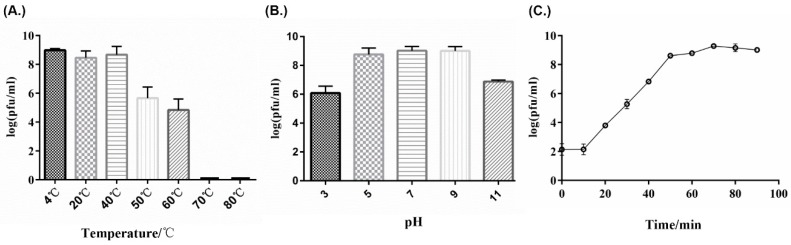
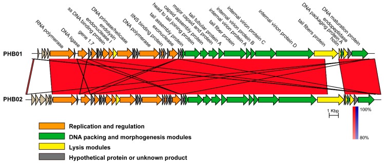
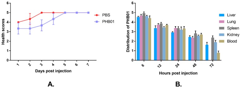
A lytic bacteriophage PHB01 specific for Pasteurella multocida type D was isolated from the sewage water collected from a pig farm. This phage had the typical morphology of the family Podoviridae, order Caudovirales, presenting an isometric polyhedral head and a short noncontractile tail. PHB01 was able to infect most of the non-toxigenic P. multocida type D strains tested, but not toxigenic type D strains and those belonging to other capsular types. Phage PHB01, the first lytic phage specific for P. multocida type D sequenced thus far, presents a 37,287-bp double-stranded DNA genome with a 223-bp terminal redundancy. The PHB01 genome showed the highest homology with that of PHB02, a lytic phage specific for P. multocida type A. Phylogenetic analysis showed that PHB01 and PHB02 were composed of a genus that was close to the T7-virus genus. In vivo tests using mouse models showed that the administration of PHB01 was safe to the mice and had a good effect on treating the mice infected with different P. multocida type D strains including virulent strain HN05. These findings suggest that PHB01 has a potential use in therapy against infections caused by P. multocida type D.

Keywords: P. multocida type D; bacteriophage; isolation; lytic; therapeutic application.


Conflict of interest statement

The authors declare no conflict of interest.

Figures

Figure 1
Figure 1
Experimental scheme for the evaluation of PHB01 treatment efficacy in mice infected with P. multocida type D. Each of the mice in the P. multocida-challenged groups were given a treatment of PHB01 at 108 PFU (Groups II and V) or PBS (Groups I and IV) at 6 h following bacterial inoculation, and then twice a day for five days.
Figure 2
Figure 2
Morphological characteristics of phage PHB01. (A) Plaques of phage PHB01 on Pasteurella multocida HND065; (B) Transmission electron micrograph of phage PHB01 (marked with white arrows).
Figure 3
Figure 3
Biological properties of phage PHB01. (A) Sensitivity to temperature variations; (B) Sensitivity to pH variations; (C) One-step growth curve.
Figure 4
Figure 4
Phylogenetic tree analysis based on the alignments of amino acid sequences of the major capsid protein ((A); Protein ID: ASD51044.1), the DNA polymerases ((B); Protein ID: ASD51034.1), and the RNA polymerases ((C); Protein ID: ASD51019.1) of the Autographivirinae subfamily phages from GenBank. Phage PHB01 is indicated by the black circle. The evolutionary trees were constructed using the neighbor-joining method with the Poisson correction model. One thousand bootstrap repetitions were performed. Sequences were aligned by the ClustalW package carried by MEGAX [35].
Figure 5
Figure 5
A co-linearity comparison diagram of the genomic organization at the nucleotide level between Pasteurella phages PHB01 and PHB02. The figure was generated via Easyfig v.2.0. The color code refers to the BLASTn identity of those regions between genomes. Arrows represent putative CDSs encoded by different genomes.
Figure 6
Figure 6
Safety test of PHB01 on mice. (A) Health scores given to the mice in different groups; (B) Distribution of phage PHB01 on the main organs of the mice. Data are presented as the mean ± SD.
Figure 7
Figure 7
Histopathological analysis of the main organs collected from the mice received a challenge of phage PHB01 and/or PBS. The liver, spleen, kidney, and lungs were fixed with 4% formalin. Tissue sections were stained with hematoxylin and eosin or toluidine blue.
Figure 8
Figure 8
Protective effects of the phage PHB01 in mice challenged with wild-type P. multocida strain HND065 and HN05. (A) Health scores given to the mice in different groups. The total score for the health status of each group was recorded at least three times per day. The data are expressed as the mean ± SD; (B) Survival curve of the mice in each of the groups during the experiment.

Similar articles

Cited by

References

    1. Carter G.R. Studies on Pasteurella multocida. I. A hemagglutination test for the identification of serological types. Am. J. Vet. Res. 1955;16:481–484. - PubMed
    1. Wilkie I.W., Harper M., Boyce J.D., Adler B. Pasteurella multocida: Diseases and pathogenesis. Curr. Top. Microbiol. Immunol. 2012;361:1–22. doi: 10.1007/82_2012_216. - DOI - PubMed
    1. Katechakis N., Maraki S., Dramitinou I., Marolachaki E., Koutla C., Ioannidou E. An unusual case of Pasteurella multocida bacteremic meningitis. J. Infect. Public Health. 2018 doi: 10.1016/j.jiph.2018.05.012. - DOI - PubMed
    1. Abreu F., Rodriguez-Lucas C., Rodicio M.R., Vela A.I., Fernandez-Garayzabal J.F., Leiva P.S., Cuesta F., Cid D., Fernandez J. Human Pasteurella multocida Infection with Likely Zoonotic Transmission from a Pet Dog, Spain. Emerg. Infect. Dis. 2018;24:1145–1146. doi: 10.3201/eid2406.171998. - DOI - PMC - PubMed
    1. Pak S., Valencia D., Decker J., Valencia V., Askaroglu Y. Pasteurella multocida pneumonia in an immunocompetent patient: Case report and systematic review of literature. Lung India. 2018;35:237–240. doi: 10.4103/lungindia.lungindia_482_17. - DOI - PMC - PubMed

Publication types

LinkOut - more resources