Review
. 2019 Jan 21;8(1):77.
doi: 10.3390/cells8010077.

Roles of Autophagy-Related Genes in the Pathogenesis of Inflammatory Bowel Disease

Affiliations
Review

Roles of Autophagy-Related Genes in the Pathogenesis of Inflammatory Bowel Disease

Sup Kim et al. Cells. .

Abstract

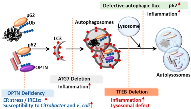
Autophagy is an intracellular catabolic process that is essential for a variety of cellular responses. Due to its role in the maintenance of biological homeostasis in conditions of stress, dysregulation or disruption of autophagy may be linked to human diseases such as inflammatory bowel disease (IBD). IBD is a complicated inflammatory colitis disorder; Crohn's disease and ulcerative colitis are the principal types. Genetic studies have shown the clinical relevance of several autophagy-related genes (ATGs) in the pathogenesis of IBD. Additionally, recent studies using conditional knockout mice have led to a comprehensive understanding of ATGs that affect intestinal inflammation, Paneth cell abnormality and enteric pathogenic infection during colitis. In this review, we discuss the various ATGs involved in macroautophagy and selective autophagy, including ATG16L1, IRGM, LRRK2, ATG7, p62, optineurin and TFEB in the maintenance of intestinal homeostasis. Although advances have been made regarding the involvement of ATGs in maintaining intestinal homeostasis, determining the precise contribution of autophagy has remained elusive. Recent efforts based on direct targeting of ATGs and autophagy will further facilitate the development of new therapeutic opportunities for IBD.

Keywords: ATGs; autophagy; inflammatory bowel diseases; intestinal homeostasis.


Conflict of interest statement

The authors declare no conflict of interest.

Figures

Figure 1
Figure 1
Overview of autophagy, selective autophagy and ATGs (A) Molecular machinery of autophagy process. After mTOR inhibition or AMPK activation, the autophagy process begins with the biogenesis of the phagophore/isolation membrane. The ATG16L1-ATG5-ATG12 and LC3-II-PE conjugates participate in autophagosome formation process. The mature autophagosomes are fused with a late endosome and lysosome to initiate degradation of cargos. Finally, cells recycle the released products in cytosol. (B) Selective autophagy clears various targets such as subcellular structure, bacteria, protein and lipid aggregates.
Figure 2
Figure 2
The process of autophagy flux and involved genes. The autophagy flux is depicted. A normal autophagic flux includes the autophagosome formation and maturation step and the autolysosome formation step. The possible conditions associated with involved genes are depicted: (1) OPTN deficiency leads to an accumulation of IRE1α and increased susceptibility of Citrobacter and E. coli. (2) ATG7 deletion is associated with increased inflammation. (3) TFEB deletion results in increased inflammation and lysosomal defect.
Figure 3
Figure 3
Summary diagram showing the role of ATG16L1 in the Crohn’s disease. The left panel demonstrates the normal host defense mechanism against intracellular pathogens. Healthy cells exhibit normal granule secretion, autophagic activity, ER stress response and permeability. The right panel shows the ATG16L1 T300A variant cells defective in granule secretion, autophagy process, IRE1α degradation and tight junction barrier function.
Figure 4
Figure 4
Autophagy targets adherent-invasive Escherichia coli (AIEC), Mycobacteria, Salmonella and Listeria by different mechanisms. Stimulation with IFN-γ induce IRGM to clear intracellular bacteria. Furthermore, IRGM can be induced by IFN-γ contribute to cell-autonomous defense though autophagy activation via the recruitment of both autophagic and SNARE adaptor proteins during infection.
Figure 5
Figure 5
The pathogenic roles of LRRK in non-intestine and intestine diseases. In Parkinson’s disease, LRRK2 is involved in cell cycle/survival, ROS generation, autophagolysosomal pathway, mitophagy and inflammation. LRRK2 plays a key role in intestine homeostasis through regulating NFAT, NF-κB, ROS generation, inflammation and autophagy.

References

    1. Ke P., Shao B.Z., Xu Z.Q., Chen X.W., Liu C. Intestinal Autophagy and Its Pharmacological Control in Inflammatory Bowel Disease. Front. Immunol. 2016;7:695. doi: 10.3389/fimmu.2016.00695. - DOI - PMC - PubMed
    1. Podolsky D.K. Inflammatory bowel disease. N. Engl. J. Med. 2002;347:417–429. doi: 10.1056/NEJMra020831. - DOI - PubMed
    1. Xavier R.J., Podolsky D.K. Unravelling the pathogenesis of inflammatory bowel disease. Nature. 2007;448:427–434. doi: 10.1038/nature06005. - DOI - PubMed
    1. Muzes G., Tulassay Z., Sipos F. Interplay of autophagy and innate immunity in Crohn’s disease: A key immunobiologic feature. World J. Gastroenterol. 2013;19:4447–4454. doi: 10.3748/wjg.v19.i28.4447. - DOI - PMC - PubMed
    1. Fritz T., Niederreiter L., Adolph T., Blumberg R.S., Kaser A. Crohn’s disease: NOD2, autophagy and ER stress converge. Gut. 2011;60:1580–1588. doi: 10.1136/gut.2009.206466. - DOI - PMC - PubMed

Publication types

Substances

LinkOut - more resources