. 2019 Jan 22;14(1):6.
doi: 10.1186/s13024-018-0303-3.

Inhibition of monocyte-like cell extravasation protects from neurodegeneration in DBA/2J glaucoma

Affiliations

Inhibition of monocyte-like cell extravasation protects from neurodegeneration in DBA/2J glaucoma

Pete A Williams et al. Mol Neurodegener. .

Abstract

Background: Glaucoma is characterized by the progressive dysfunction and loss of retinal ganglion cells. Recent work in animal models suggests that a critical neuroinflammatory event damages retinal ganglion cell axons in the optic nerve head during ocular hypertensive injury. We previously demonstrated that monocyte-like cells enter the optic nerve head in an ocular hypertensive mouse model of glaucoma (DBA/2 J), but their roles, if any, in mediating axon damage remain unclear.

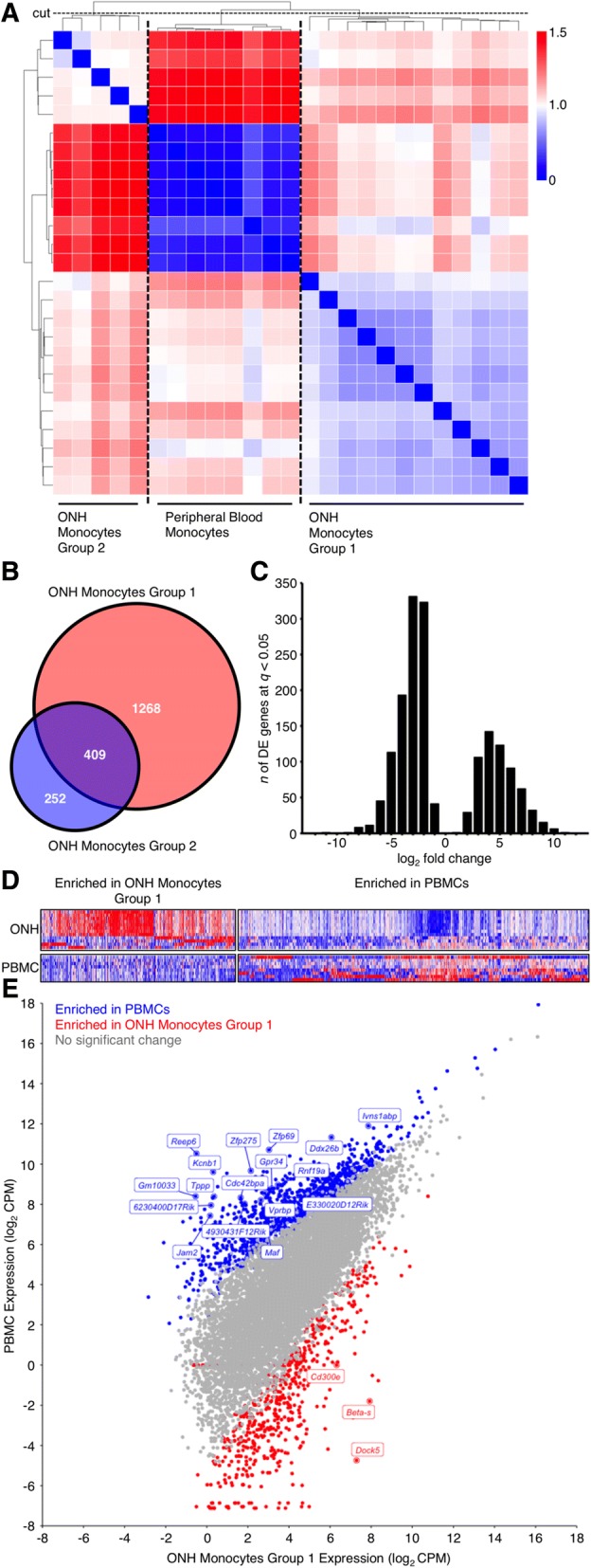
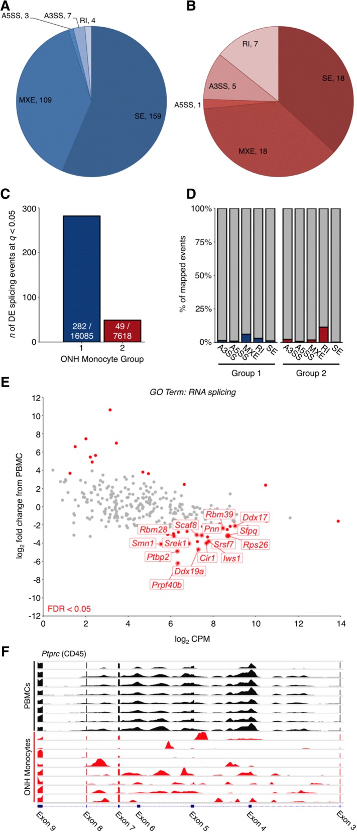
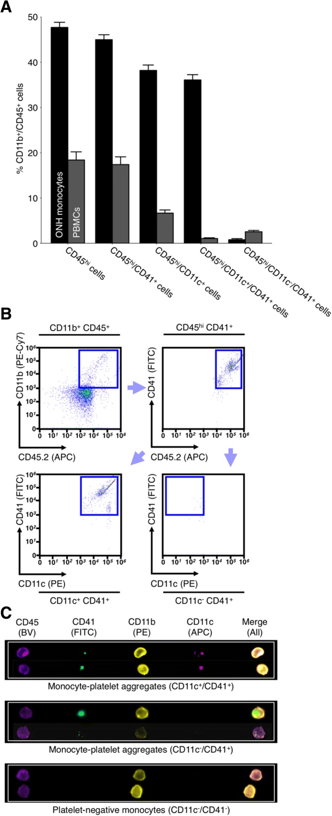
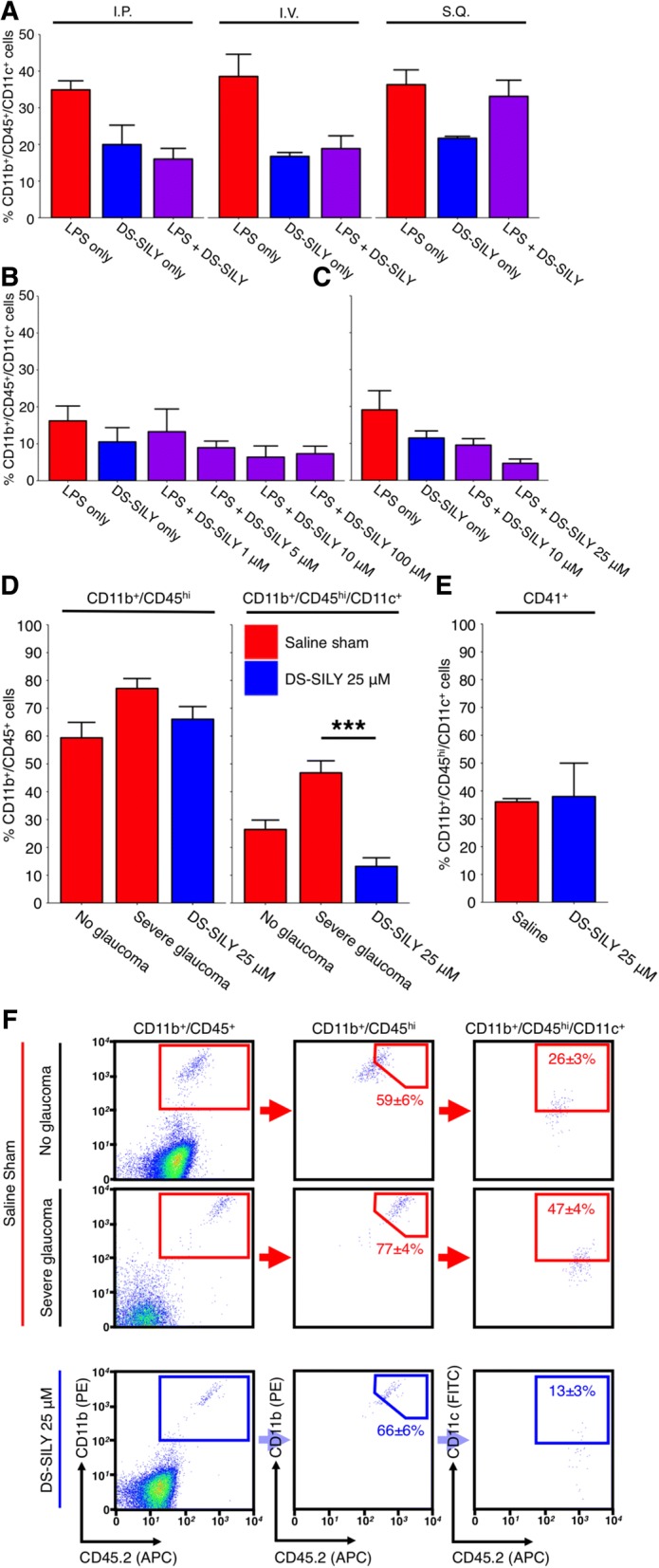
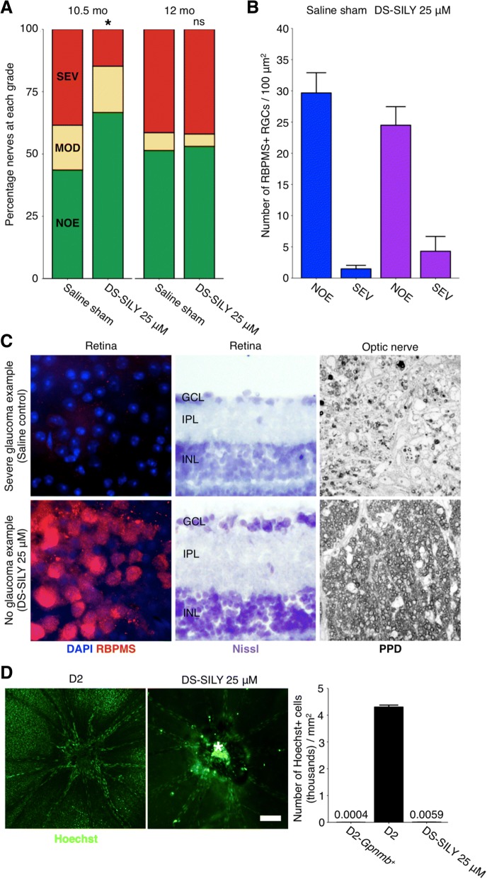
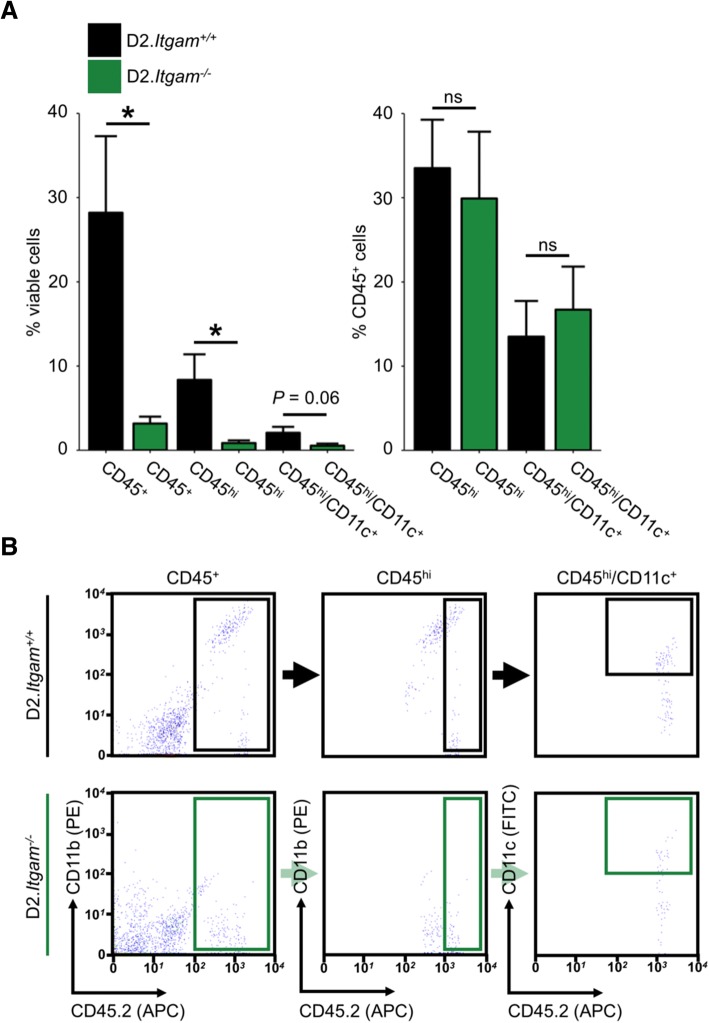
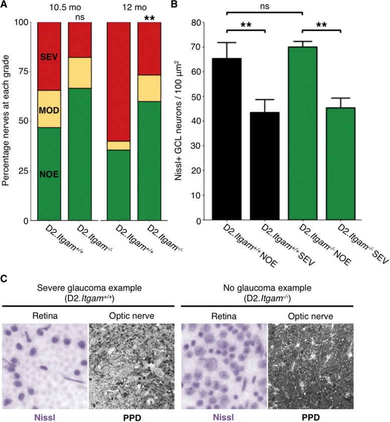
Methods: To understand the function of these infiltrating monocyte-like cells, we used RNA-sequencing to profile their transcriptomes. Based on their pro-inflammatory molecular signatures, we hypothesized and confirmed that monocyte-platelet interactions occur in glaucomatous tissue. Furthermore, to test monocyte function we used two approaches to inhibit their entry into the optic nerve head: (1) treatment with DS-SILY, a peptidoglycan that acts as a barrier to platelet adhesion to the vessel wall and to monocytes, and (2) genetic targeting of Itgam (CD11b, an immune cell receptor that enables immune cell extravasation).

Results: Monocyte specific RNA-sequencing identified novel neuroinflammatory pathways early in glaucoma pathogenesis. Targeting these processes pharmacologically (DS-SILY) or genetically (Itgam / CD11b knockout) reduced monocyte entry and provided neuroprotection in DBA/2 J eyes.

Conclusions: These data demonstrate a key role of monocyte-like cell extravasation in glaucoma and demonstrate that modulating neuroinflammatory processes can significantly lessen optic nerve injury.

Keywords: Extravasation; Glaucoma; Monocyte; Neuroinflammation; Optic nerve; Platelet; RNA-sequencing; Retinal ganglion cell; Vascular leakage.


Conflict of interest statement

Ethics approval

All breeding and experimental procedures were undertaken in accordance with the Association for Research for Vision and Ophthalmology Statement for the Use of Animals in Ophthalmic and Research. The Institutional Biosafety Committee (IBC) and the Animal Care and Use Committee (ACUC) at The Jackson Laboratory approved this study.

Consent for publication

N/A.

Competing interests

The authors declare that they have no competing interests.

Publisher’s Note

Springer Nature remains neutral with regard to jurisdictional claims in published maps and institutional affiliations.

Figures

Fig. 1
Fig. 1
RNA-sequencing of monocytes in glaucoma. a Heatmap correlations of all samples (Spearman’s rho, blue = high correlation, red = lowest correlation). Dendrogram is shown in grey at top and left of heatmap. The cut-off to generate clusters on the dendrogram is shown as the horizontal dashed line labelled “cut”. Cutting strategy was defined as all control (peripheral blood monocytes; PBMC) samples falling into one group with the maximum number of total samples retained. b Venn diagram showing number of DE genes (FDR, q < 0.05) between ONH Monocytes Group 1 vs. PBMCs and ONH Monocytes Group 2 vs. PBMCs (red and blue), and shared DE genes between groups (purple). c Histogram of DE genes by fold change between ONH Monocytes Group 1 vs. PBMCs. d Heatmap showing all DE genes (x) by sample (y). Red = highly expressed, blue = lowly expressed. e Scatter plot showing all genes (dots, not DE = grey, DE and enriched in PBMCs = blue, DE and enriched in ONH Monocytes Group 1 = red). Top 20 DE genes are named
Fig. 2
Fig. 2
RNA alternate splicing in ONH monocytes. RNA alternative splicing analysis was performed using MATS [42]. a and b Pie diagrams showing number of differentially expressed splicing events (FDR < 0.05) between ONH Monocytes Group 1 (a) and ONH Monocytes Group 2 (b) vs. PBMCs. Events were classed as either A3SS (alternate 3’ SS), A5SS (alternate 5’ SS), RI (retained introns), SE (skipped exons), or MXE (mutually exclusive exons). c Total number of DE splicing events / total number of mapped splicing events. d Percentage of DE splicing events. e Scatter plot for all genes under the GO Term: RNA splicing (GO:0006395) in ONH Monocytes Group 1 vs. PBMCs. Grey = not DE, red = DE (FDR < 0.05). DE genes at FDR < 0.01 with a log2 CPM > 5 are named. f Transcriptome plot (IGV) showing alternative splicing (skipping of exons 4, 5, and 6) of Ptprc (CD45). Black = PBMC representative samples, red = ONH monocyte representative samples
Fig. 3
Fig. 3
The majority of monocytes entering ONH tissue during glaucomatous progression are platelet bound. We have previously demonstrated that monocytes entering the ONH tissue during glaucoma are CD45hi/CD11b+/CD11c+ (Additional file 1: Figure S1 and [9]). RNA-sequencing analysis of ONH monocytes implicates platelets early in glaucoma pathogenesis. To determine if monocyte-platelet aggregates were present in the ONH in glaucoma flow cytometry was performed. a and b Antibodies against CD41 (a platelet marker) demonstrated that the majority (> 90%) of CD45hi/CD11b+/CD11c+ ONH monocytes were also platelet bound (n = 18 ONHs) compared to < 2% of PBMCs (n = 10 peripheral blood samples). c When visualized using imaging flow cytometry there were evident platelets bound to monocytes in the ONH (green, c) (n = 4 ONHs). Examples of monocyte-platelet aggregates and platelet negative monocytes are shown. BV = brilliant violet (blue/violet), FITC = fluorescein isothiocyanate (green), PE = phycoerythrin (yellow/orange), APC = allophycocyanin (red)
Fig. 4
Fig. 4
DS-SILY prevents monocyte entry to the ONH. a, b and c 1 μg/g [body weight] LPS delivered by intraperitoneal injection is sufficient to drive monocyte entry into the ONH of young, non-diseased B6 mice after 21 days (a, red bars). To prevent monocyte entry into the ONH DS-SILY was administered by three different routes; DS-SILY was protective when administered by an intravenous route, (a, purple bars) in a dose dependent manner (b). Although administration by IP had similar potency to IV, its effects were less long-lasting, lasting; IV lasted > 21 days; IP only 3–5 days (not shown) (n = 4 for all route testing conditions, n > 6 for all concentration testing conditions). DS-SILY administered at 1 μM was used as a sham control (blue bars in a, b and c). c Further testing in young D2 mice determined that DS-SILY was most potent at 25 μM (n = 11 LPS only; 9 DS-SILY only; 23 DS-SILY 10 μM; 8 DS-SILY 25 μM). d Subsequently 25 μM DS-SILY was administered every 21 days via intravenous injection to the tail vein of D2 mice starting at 6–7 mo of age. Mice administered DS-SILY and saline sham controls were harvested at 10.5 mo and ONHs assessed for monocyte entry by flow cytometry. DS-SILY robustly protected from CD45hi/CD11b+/CD11c+ monocyte entry (n = 24 saline; 23 DS-SILY). e A subset of monocytes that still entered in DS-SILY treated ONHs were still platelet positive (n = 2 pools of 4 ONHs each). f Example flow plots are shown for (d). CD11b+/CD45+ = myeloid-derived cells (i.e. microglia and monocytes in the ONH), CD11b+/CD45hi = all monocytes, CD11b+/CD45hi/CD11c+ = infiltrating monocyte-like cells, CD11b+/CD45hi/CD11c+/CD41+ = infiltrating monocyte-like cells that are platelet bound (i.e. monocyte-platelet aggregates)
Fig. 5
Fig. 5
DS-SILY protects from D2 glaucoma at 10.5 mo of age. 25 μM DS-SILY was administered every 21 days via intravenous injection to the tail vein of D2 mice starting at 6–7 mo of age. Mice administered DS-SILY and saline sham controls were harvested at 10.5 and 12 mo and optic nerves and retinas were assessed for glaucomatous neurodegeneration. a DS-SILY administration significantly protected D2 eyes from glaucoma at 10.5 mo as assessed by optic nerve assessment (green = NOE; no detectable glaucoma but called no or early glaucoma as some eyes have early gene expression changes, yellow = MOD; moderate glaucoma, red = SEV; severe glaucoma). b Agreeing with this, RGC numbers were preserved in protected eyes with no detectable glaucoma (blue bars = saline sham, purple bars = DS-SILY). Eyes with severe glaucoma had also lost their RGCs indicating that DS-SILY treatment did not uncouple somal and axonal degeneration (SEV bars)). c Examples of no glaucoma and severe glaucoma retinas as assessed by RBPMS staining (an RGC marker; left; n = 5 for each condition), Nissl staining (center; n = 5 for each condition), and optic nerve cross sections assessed by PPD staining (right; n = 55 saline 10.5 mo; 64 saline 12 mo; 80 DS-SILY 10.5 mo; 66 DS-SILY 12 mo). Top row shows examples of severe retinas from representative eyes with severe optic nerve damage (SEV) as assessed by PPD staining of the optic nerve, bottom row shows examples of retinas with no neurodegeneration in the optic nerve (NOE graded nerves). d DS-SILY administration potently prevents vascular leakage in the retina at 9–9.5 mo (n = 18). Vascular leakage was assessed by an intravenous injection of the nuclear label Hoechst (green). Retinas were flat-mounted and Hoechst positive ganglion cell layer nuclei (excluding vascular endothelial cell nuclei) were counted across the retina from 8 representative regions in each eye. The bright region (*) over the optic nerve head in the right hand panel is due to labelling of cells in a residual tuft of hyaloid vasculature over the ONH (as often occurs in mice) and does not indicate increased leakage in the optic nerve. Scale bar = 100 μm. The numbers above D2-Gpnmb+ and DS-SILY 25 μM represent the data points
Fig. 6
Fig. 6
Genetic ablation of CD11b (Itgam) decreases monocyte number in the ONH. To determine whether genetic ablation of Itgam affects monocyte entry into the ONH during glaucoma, D2.Itgam−/− mice and genetic controls were harvested at 10.5 mo and ONHs assessed for monocytes by flow cytometry. a CD45+ and CD45hi cell number in the ONH was significantly decreased in Itgam−/− mice (a, left). Homozygous knockout mice lack CD11b and so the gating strategy compared to % viable cells. The relative proportions of CD45hi and CD45hi/CD11c+ types of myeloid derived cells were not changed (a, right) (n = 12 (D2.Itgam+/+), 10 (D2.Itgam−/−)). b Example flow plots and gating strategy
Fig. 7
Fig. 7
Genetic ablation of CD11b (Itgam) protects from glaucoma to aged time points. a D2.Itgam−/− mice are significantly protected from glaucoma as determined by optic nerve assessment (green = NOE; no detectable glaucoma but called no or early glaucoma as some of these eyes have early gene expression changes, yellow = MOD; moderate glaucoma, red = SEV; severe glaucoma). b Agreeing with this, RGC numbers were preserved in eyes with no detectable glaucoma. Eyes with severe glaucoma had also lost their RGCs indicating that loss of CD11b did not uncouple somal and axonal degeneration. c Example optic nerves and retinas. Left shows examples of severe retinas from eyes with severe optic nerve damage (SEV) as assessed by PPD staining of the optic nerve, right shows examples of retinas with no neurodegeneration in the optic nerve (NOE graded nerves). (Soma counts, n = 6 for each condition; optic nerve analysis as assessed by PPD staining (right) n = 32 D2.Itgam+/+ 10.5 mo; 45 D2.Itgam+/+ 12 mo; 45 D2.Itgam−/− 10.5 mo; 45 D2.Itgam−/− 12 mo)

References

    1. Quigley HA, Broman AT. The number of people with glaucoma worldwide in 2010 and 2020. Br J Ophthalmol. 2006;90:262–267. doi: 10.1136/bjo.2005.081224. - DOI - PMC - PubMed
    1. Tham YC, Li X, Wong TY, Quigley HA, Aung T, Cheng CY. Global prevalence of glaucoma and projections of glaucoma burden through 2040: a systematic review and meta-analysis. Ophthalmology. 2014;121:2081–2090. doi: 10.1016/j.ophtha.2014.05.013. - DOI - PubMed
    1. Williams PA, Marsh-Armstrong N, Howell GR, Participants LIIoAaGN Neuroinflammation in glaucoma: a new opportunity. Exp Eye Res. 2017;157:20–27. doi: 10.1016/j.exer.2017.02.014. - DOI - PMC - PubMed
    1. Margeta MA, Lad EM, Proia AD. CD163+ macrophages infiltrate axon bundles of postmortem optic nerves with glaucoma. Graefes Arch Clin Exp Ophthalmol. 2018;256(12):2449–2456. doi: 10.1007/s00417-018-4081-y. - DOI - PMC - PubMed
    1. Harder JM, Braine CE, Williams PA, Zhu X, MacNicoll KH, Sousa GL, Buchanan RA, Smith RS, Libby RT, Howell GR, John SWM. Early immune responses are independent of RGC dysfunction in glaucoma with complement component C3 being protective. Proc Natl Acad Sci U S A. 2017;114(19):E3839–E3848. doi: 10.1073/pnas.1608769114. - DOI - PMC - PubMed

Publication types