. 2019 Jan 22;14(1):7.
doi: 10.1186/s13024-019-0307-7.

Reduced presynaptic vesicle stores mediate cellular and network plasticity defects in an early-stage mouse model of Alzheimer's disease

Affiliations

Reduced presynaptic vesicle stores mediate cellular and network plasticity defects in an early-stage mouse model of Alzheimer's disease

Shreaya Chakroborty et al. Mol Neurodegener. .

Abstract

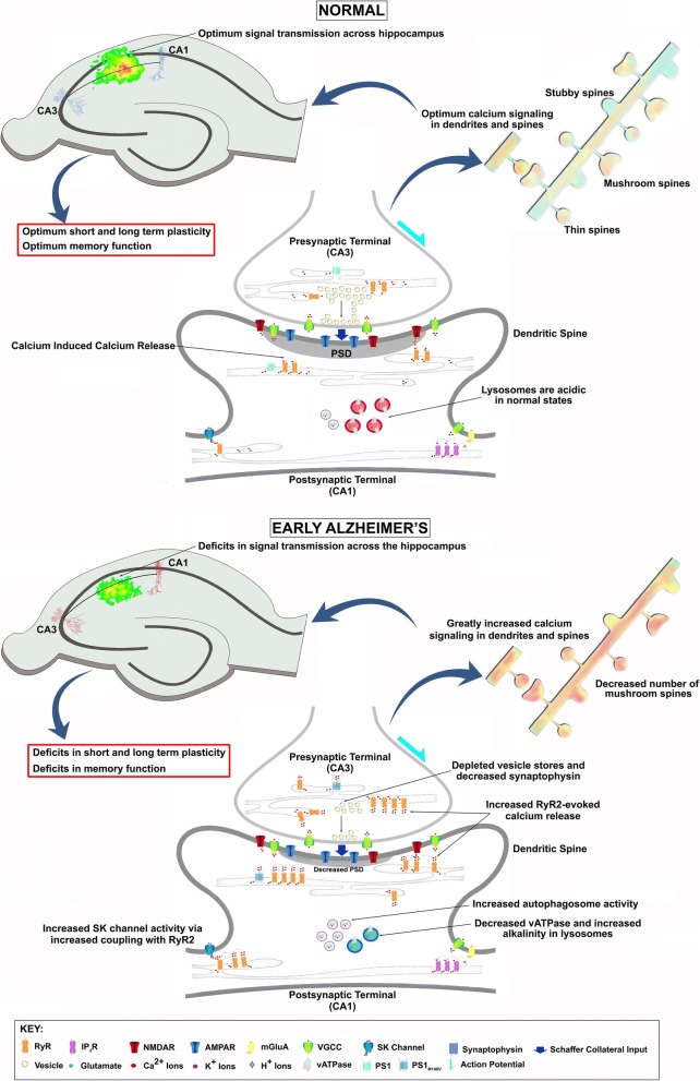
Background: Identifying effective strategies to prevent memory loss in AD has eluded researchers to date, and likely reflects insufficient understanding of early pathogenic mechanisms directly affecting memory encoding. As synaptic loss best correlates with memory loss in AD, refocusing efforts to identify factors driving synaptic impairments may provide the critical insight needed to advance the field. In this study, we reveal a previously undescribed cascade of events underlying pre and postsynaptic hippocampal signaling deficits linked to cognitive decline in AD. These profound alterations in synaptic plasticity, intracellular Ca2+ signaling, and network propagation are observed in 3-4 month old 3xTg-AD mice, an age which does not yet show overt histopathology or major behavioral deficits.

Methods: In this study, we examined hippocampal synaptic structure and function from the ultrastructural level to the network level using a range of techniques including electron microscopy (EM), patch clamp and field potential electrophysiology, synaptic immunolabeling, spine morphology analyses, 2-photon Ca2+ imaging, and voltage-sensitive dye-based imaging of hippocampal network function in 3-4 month old 3xTg-AD and age/background strain control mice.

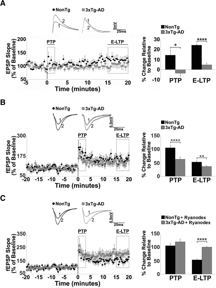
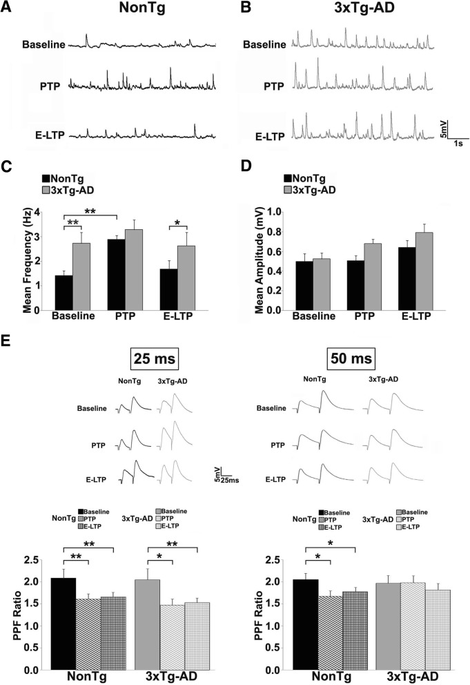
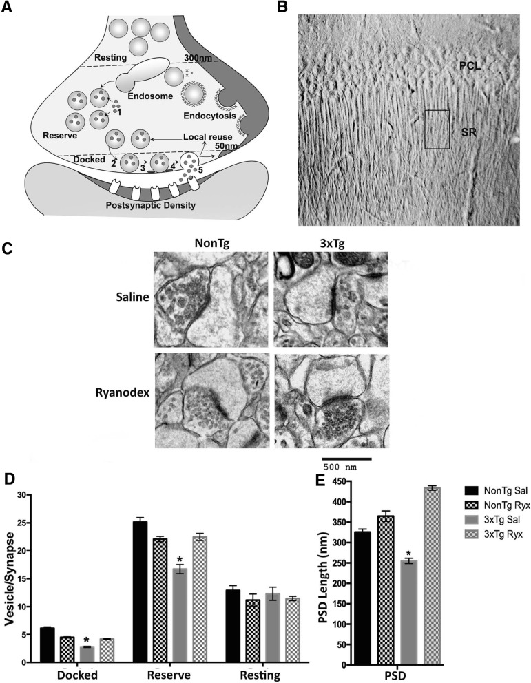
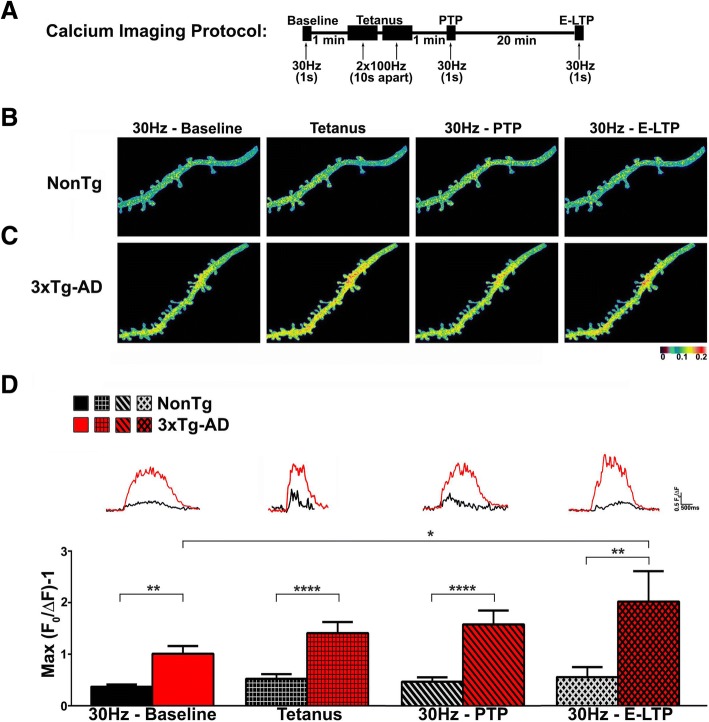
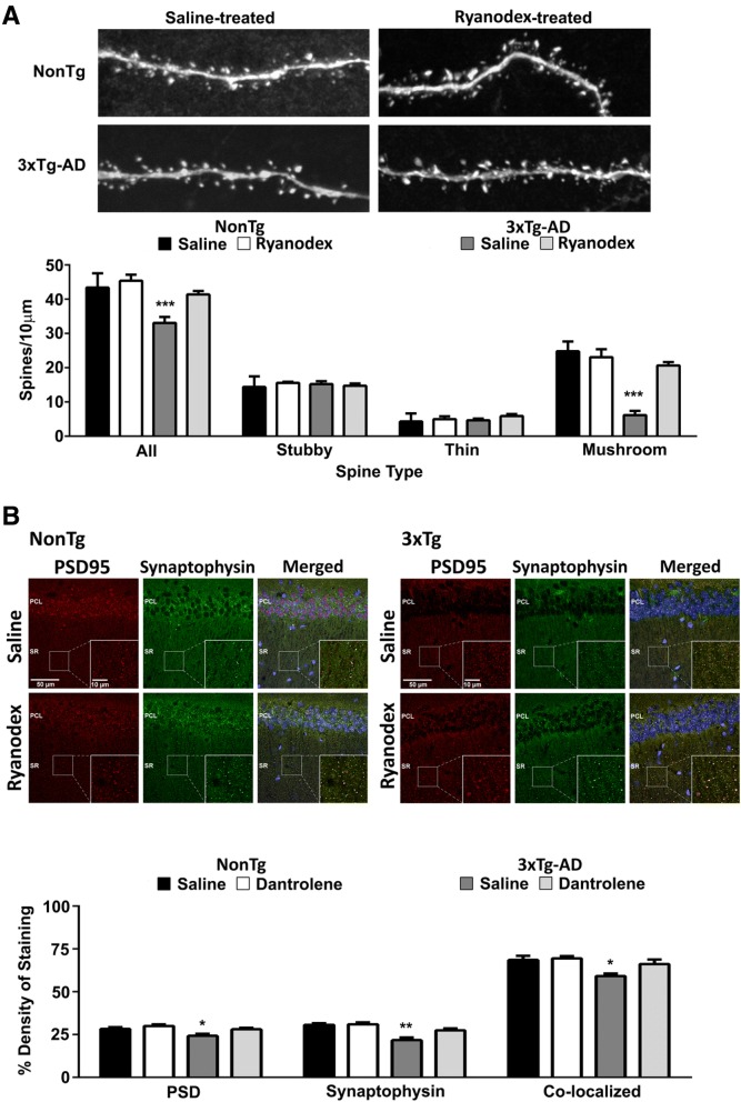
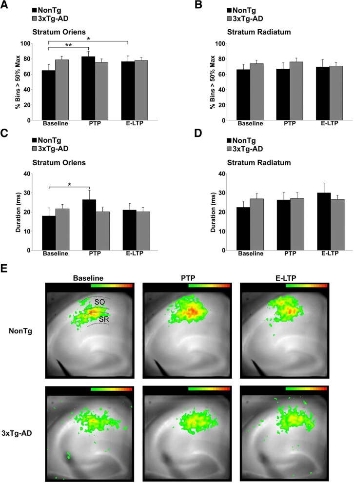
Results: In 3xTg-AD mice, short-term plasticity at the CA1-CA3 Schaffer collateral synapse is profoundly impaired; this has broader implications for setting long-term plasticity thresholds. Alterations in spontaneous vesicle release and paired-pulse facilitation implicated presynaptic signaling abnormalities, and EM analysis revealed a reduction in the ready-releasable and reserve pools of presynaptic vesicles in CA3 terminals; this is an entirely new finding in the field. Concurrently, increased synaptically-evoked Ca2+ in CA1 spines triggered by LTP-inducing tetani is further enhanced during PTP and E-LTP epochs, and is accompanied by impaired synaptic structure and spine morphology. Notably, vesicle stores, synaptic structure and short-term plasticity are restored by normalizing intracellular Ca2+ signaling in the AD mice.

Conclusions: These findings suggest the Ca2+ dyshomeostasis within synaptic compartments has an early and fundamental role in driving synaptic pathophysiology in early stages of AD, and may thus reflect a foundational disease feature driving later cognitive impairment. The overall significance is the identification of previously unidentified defects in pre and postsynaptic compartments affecting synaptic vesicle stores, synaptic plasticity, and network propagation, which directly impact memory encoding.

Keywords: 2-photon imaging; Alzheimer’s disease; Calcium; Electron microscopy; Hippocampus; Mouse model; Network imaging; Patch clamp; Ryanodine receptor; Short-term plasticity; Spines; Synaptic; Synaptic vesicles.


Conflict of interest statement

Ethics approval and consent to participate

Not applicable.

Consent for publication

Not applicable.

Competing interests

The authors declare that they have no competing interests.

Publisher’s Note

Springer Nature remains neutral with regard to jurisdictional claims in published maps and institutional affiliations.

Figures

Fig. 1
Fig. 1
Short term plasticity deficits in 3xTg-AD CA1 region reflect RyR-Ca2+ abnormalities. a-c Left: Graphs show averaged time course of Schaffer collateral-evoked synaptic responses from NonTg (black circles) and 3xTg-AD (gray squares) in the hippocampal region using (a) whole cell patch clamp from CA1 pyramidal neurons (NonTg n = 10 neurons/6 mice; 3xTg-AD n = 10 neurons/6 mice) and (b-c) extracellular field potential recordings in CA1 stratum radiatum (NonTg n = 8 slices/5 mice; 3xTg-AD n = 8 slices/5 mice). Right: Bar graphs show averaged % change over baseline in the evoked response post-tetanus in 3xTg-AD (gray) compared to NonTg (black). Inset: Representative traces [1] before and [2] after tetanus from NonTg (black) and 3xTg-AD (gray) neurons. PTP and E-LTP are impaired in (a) individual neurons and (b) field potentials of 3xTg-AD CA1 neurons compared to NonTg neurons. c 30-day Ryanodex treatment restores or enhances PTP and E-LTP in 3xTg-AD CA1 circuits (n = 5 slices/5 mice; NonTg n = 5 slices/5 mice) . The arrow denotes the time of tetanus. Data are presented as Mean ± SEM; *p < 0.05, **p < 0.01 and ****p < 0.0001 represents significantly different from NonTg
Fig. 2
Fig. 2
Impaired spontaneous and evoked presynaptic release properties in 3xTg-AD mice. Representative traces of sEPSPs from (a) NonTg (black) and (b) 3xTg-AD (gray) CA1 neurons at baseline, PTP, and E-LTP time-points. c Bar graphs show elevated averaged sEPSP frequency in 3xTg-AD (gray) neurons compared to NonTg (black) at baseline, and remains elevated during PTP and E-LTP. In NonTg neurons, sEPSP frequency is increased during PTP relative to its baseline, and returns to baseline during E-LTP. d sEPSP amplitude is not significantly different between NonTg and 3xTg-AD neurons at all time-points. Data are presented as Mean ± SEM; *p < 0.05 and **p < 0.01 represents significantly different from NonTg, **^p < 0.01 represents significantly different from pre-tetanus baseline. e Bar graphs show averaged paired pulse ratio (PPR) responses during baseline, PTP and E-LTP at 25 ms ISI (left) and 50 ms ISI (right) from NonTg (black, and patterned black) and 3xTg-AD neurons (gray and patterned gray). At the 50 ms ISI, the 3xTg-AD mice do not show the reduced PPR during PTP and E-LTP relative to baseline, as seen in the NonTg mice, suggesting that vesicle release probability is not further increased during these periods (NonTg n = 10 neurons/6 mice; 3xTg-AD n = 10 neurons/6 mice). Insets: Representative traces from NonTg (black) and 3xTg-AD (gray) at 25 ms and 50 ms. Data are presented as Mean ± SEM; *p < 0.05 and **p < 0.01 represent significantly different from pre-tetanus baseline
Fig. 3
Fig. 3
3xTg-AD mice have fewer synaptic vesicles in active zones and decreased PSD length. a Diagram showing synaptic vesicle cycle including [1] neurotransmitter uptake, [2] docking, [3] priming, [4] fusion and [5] release, and vesicle classification based on distance from the presynaptic membrane (docked: 0-50 nm, reserve: 50-300 nm, resting: > 300 nm) [41]. b DIC image showing region of interest within the CA1 stratum radiatum (SR) from which ultrathin sections were obtained for EM; PCL: Pyramidal Cell Layer. c Representative electron micrographs from saline- and Ryanodex-treated NonTg and 3xTg-AD asymmetric synapses (saline: NonTg n = 40 micrographs/5 mice; 3xTg-AD n = 56 micrographs/7 mice. Ryanodex treated: NonTg n = 36 micrographs/4 mice; 3xTg-AD n = 45 micrographs/5 mice). Scale bar: 500 nm, direct magnification: 30,000X, PSD: Postsynaptic Density. Bar graphs comparing (d) number of synaptic vesicles per synapse and (e) PSD length observed in NonTg and 3xTg-AD mice. Data are presented as Mean ± SEM; *p < 0.01 represents significantly different from NonTg Sal group
Fig. 4
Fig. 4
Continuously increasing synaptically-evoked Ca2+ responses in 3xTg-AD CA1 neurons during and after tetanus. a Schematic showing the synaptic stimulation and Ca2+ imaging protocol. b-c Representative pseudocolored Ca2+ images of dendritic segments from (b) NonTg and (c) 3xTg-AD neurons at baseline, tetanus, PTP and E-LTP showing increased Ca2+ responses in 3xTg-AD neurons. Colors correspond to relative Ca2+ changes indicated by the bar below. (d) Bar graphs show increased Ca2+ responses in 3xTg-AD dendrites compared to NonTg at baseline, tetanus, PTP and E-LTP (NonTg n = 10 neurons/6 mice; 3xTg-AD n = 10 neurons/6 mice). Insets: Representative traces of Ca2+ responses from NonTg (black) and 3xTg-AD (red) dendrites. Data are presented as Mean ± SEM; **p < 0.01 and ****p < 0.0001 represent significantly different from NonTg; *p < 0.05 represents significantly different from pre-tetanus baseline
Fig. 5
Fig. 5
Loss of mushroom spines and synaptic integrity is reversed by normalized RyR-Ca2+ in 3xTg-AD neurons. a Top: Images show Lucifer Yellow-filled dendrites and spines from saline- and Ryanodex-treated NonTg and 3xTg-AD CA1 neurons. Bottom: Bar graphs show a significant loss of mushroom spines in the 3xTg-AD mice, and a restoration of mushroom spine number after Ryanodex treatment in 3xTg-AD neurons. (Saline treated: NonTg n = 8 neurons/3 mice; 3xTg-AD n = 6 neurons/3 mice. Ryanodex treated: NonTg n = 4 neurons/3 mice; 3xTg-AD n = 8 neurons/3 mice; 2–5 dendritic primary or secondary branches from each cell). b Top: Confocal images (40x, single plane, axial resolution < 0.3 μm) show colocalized immunolabeling of postsynaptic PSD (postsynaptic density, red) and presynaptic (synaptophysin, green) proteins at the CA3-CA1 synapses from saline- and Ryanodex-treated NonTg and 3xTg-AD mice. Inset: higher magnification (100x) detailing synaptic labeling patterns in each condition, with yellow fluorescence indicating close localization of pre- and postsynaptic markers in the merged images. Bottom: Bar graphs show a reduction of synaptophysin and PSD95 labelling, and colocalization of pre- and postsynaptic proteins in the saline-treated 3xTg-AD mice, and that Ryanodex-treatment results in the recovery of these synaptic proteins in 3xTg-AD mice, and has no effects in the NonTg mice. (NonTg n = 36 slices/6 mice for each treatment condition; 3xTg-AD n = 36 slices/6 mice for each treatment condition). Data are presented as Mean ± SEM; *p < 0.05, **p < 0.01 and ***p < 0.001 represent significantly different from saline-treated 3xTg-AD
Fig. 6
Fig. 6
Network-level STP deficits in the CA1 SO subfield of 3xTg-AD hippocampus. Bar graphs show spread (a-b) and duration (c-d) of strong depolarizing optical signals across the CA1 subfield in response to a single pulse to CA3 at baseline, PTP and E-LTP in the SO (a, c) and SR (b, d) from NonTg (n = 9 mice, 9 slices) and 3xTg-AD mice (n = 11 mice, 11 slices). There are no changes in spread or duration of optical signals in 3xTg-AD hippocampus after tetanus, compared to increased spread and duration of optical signals in NonTg hippocampus. e Pseudocolored VSD images showing the spread of optical signals in NonTg and 3xTg-AD hippocampus during baseline, PTP and E-LTP. The SO, SP, and SR regions are indicated by dashed lines in the NonTg/Baseline image. Optical signals > 50% over baseline are shown. Data presented as Mean ± SEM; *p < 0.05 and **p < 0.01 represent significantly different from pre-tetanus baseline
Fig. 7
Fig. 7
Proposed role of RyR-Ca2+ signaling in early impairment of short term plasticity at the CA3-CA1 synapse in Alzheimer’s disease. Normal: In presynaptic CA3 terminals, RyR-evoked Ca2+-Induced-Ca2+-Release (CICR) can trigger spontaneous neurotransmitter release. During high frequency activity (such as a train of action potentials), CICR is evoked by voltage-gated Ca2+ influx to increase residual Ca2+ levels and release probability. Postsynaptically, in CA1 terminals, NMDAR-mediated Ca2+ signals are amplified by RyR-CICR in dendritic spines, a phenomenon required for plasticity induction. In other plasticity pathways, the mGluA-PLC pathway generates IP3, activating IP3R. Activation of IP3Rs produces regenerative Ca2+ waves that support plasticity and gene expression. RyR-Ca2+ also activates SK channels that modulate membrane excitability and frequency of action potential firing. Thus, optimum levels of Ca2+ signaling and synaptic plasticity proteins like synaptophysin and PSD in dendritic spines support maintenance of spine structure and signal transmission across the hippocampus, generating optimal levels of plasticity and normal memory function. Early AD: Presynaptically, increased RyR expression greatly increases CICR. Increased CICR alters vesicle cycling and cause depletion of vesicles from the readily releasable pool as well as the reserve pool. While this can be initially remedied by increasing neurotransmitter synthesis and vesicle cycling, such maladaptive mechanisms can potentially cause metabolic and oxidative stress leading to synapse loss. Postsynaptically, increased RyR-CICR can decrease PSD lengths and weaken synaptic integrity resulting in the loss of dendritic spines, specifically mushroom spines required for synaptic plasticity. Increased RyR-CICR can also increase SK channel activity which decreases neuronal excitability and increases threshold for induction of synaptic plasticity. Thus, greatly increased Ca2+ signaling and loss of proteins supporting synaptic plasticity result in the loss of synaptic integrity and dendritic mushroom spines and decreased signal transmission across the hippocampus. These results in early deficits in short and long term synaptic plasticity that ultimately causes memory impairments

References

    1. Briggs CA, Chakroborty S, Stutzmann GE. Emerging pathways driving early synaptic pathology in Alzheimer's disease. Biochem Biophys Res Commun. 2017;483(4):988–997. doi: 10.1016/j.bbrc.2016.09.088. - DOI - PMC - PubMed
    1. Chakroborty S, Stutzmann GE. Early calcium dysregulation in Alzheimer’s disease: setting the stage for synaptic dysfunction. Sci China Life Sci. 2011;54(8):752–762. doi: 10.1007/s11427-011-4205-7. - DOI - PubMed
    1. Scheff SW, Price DA, Schmitt FA, DeKosky ST, Mufson EJ. Synaptic alterations in CA1 in mild Alzheimer disease and mild cognitive impairment. Neurology. 2007;68(18):1501–1508. doi: 10.1212/01.wnl.0000260698.46517.8f. - DOI - PubMed
    1. Terry RD, Masliah E, Salmon DP, Butters N, DeTeresa R, Hill R, et al. Physical basis of cognitive alterations in Alzheimer's disease: synapse loss is the major correlate of cognitive impairment. Ann Neurol. 1991;30(4):572–580. doi: 10.1002/ana.410300410. - DOI - PubMed
    1. Marchetti C, Marie H. Hippocampal synaptic plasticity in Alzheimer's disease: what have we learned so far from transgenic models? Rev Neurosci. 2011;22(4):373–402. doi: 10.1515/rns.2011.035. - DOI - PubMed

Publication types