Case Reports
. 2019 Jan 22;7(1):4.
doi: 10.1186/s40359-019-0278-z.

The hidden identity of faces: a case of lifelong prosopagnosia

Affiliations
Case Reports

The hidden identity of faces: a case of lifelong prosopagnosia

Martin Wegrzyn et al. BMC Psychol. .

Abstract

Background: Not being able to recognize a person's face is a highly debilitating condition from which people with developmental prosopagnosia (DP) suffer their entire life. Here we describe the case of J, a 30 year old woman who reports being unable to recognize her parents, her husband, or herself in the mirror.

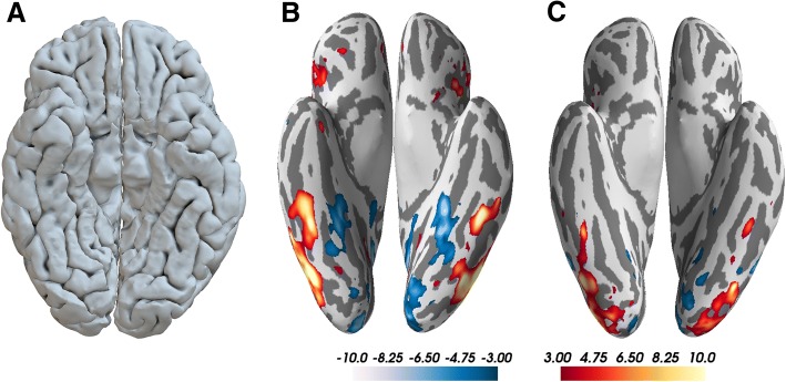
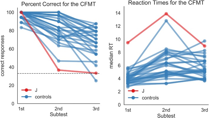
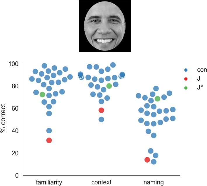
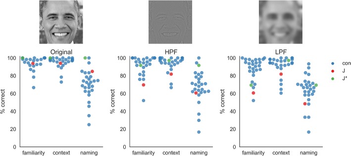
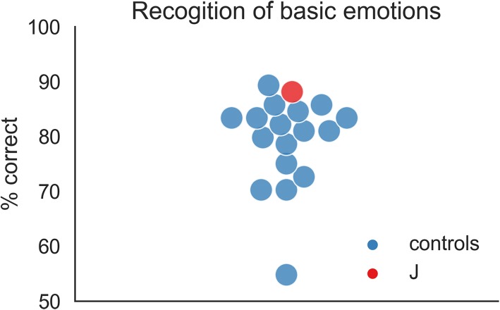
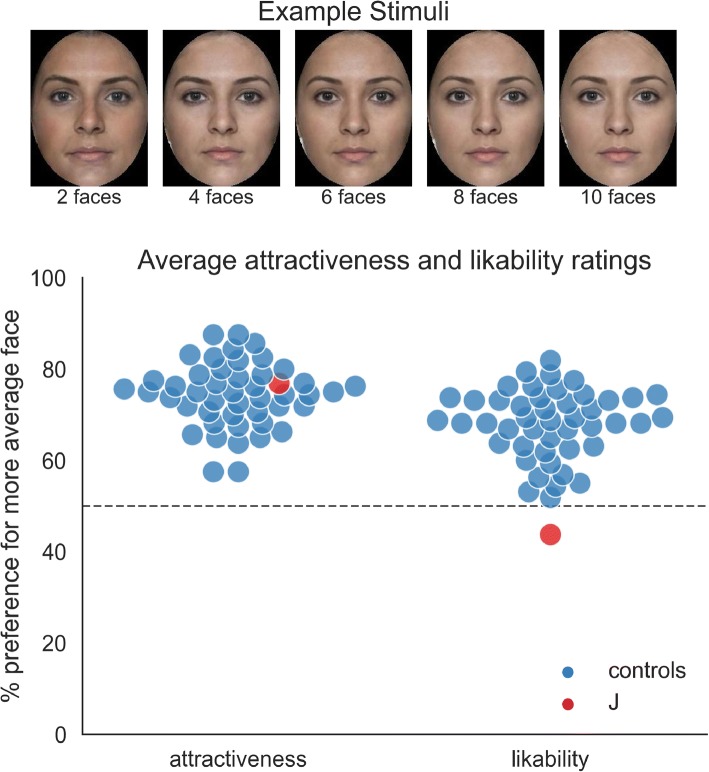
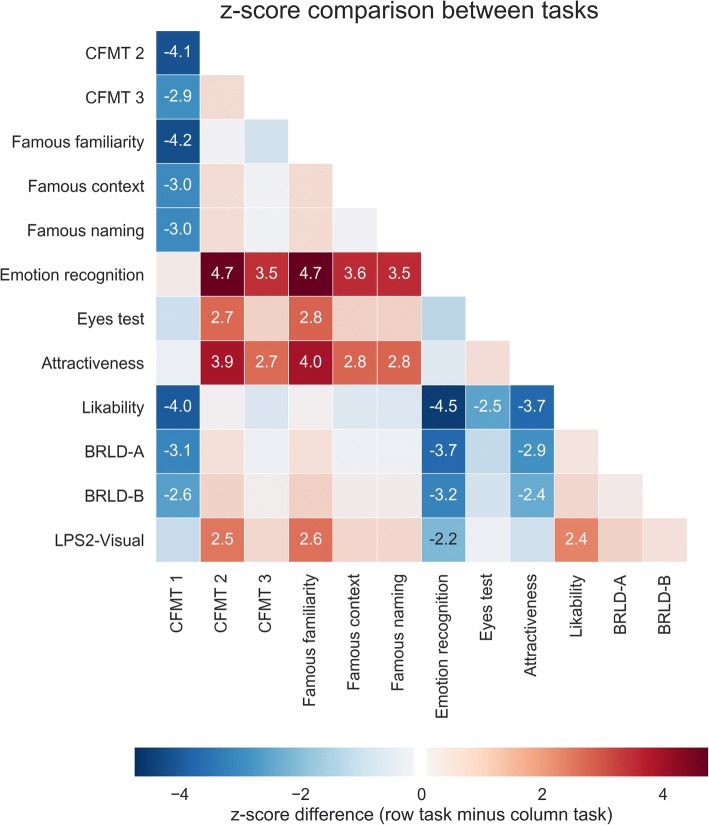
Case presentation: We set out to assess the severity of J's prosopagnosia using tests with unfamiliar as well as familiar faces and investigated whether impaired configural processing explains her deficit. To assess the specificity of the impairment, we tested J's performance when evaluating emotions, intentions, and the attractiveness and likability of faces. Detailed testing revealed typical brain activity patterns for faces and normal object recognition skills, and no evidence of any brain injury. However, compared to a group of matched controls, J showed severe deficits in learning new faces, and in recognizing familiar faces when only inner features were available. Her recognition of uncropped faces with blurred features was within the normal range, indicating preserved configural processing when peripheral features are available. J was also unimpaired when evaluating intentions and emotions in faces. In line with healthy controls, J rated more average faces as more attractive. However, she was the only one to rate them as less likable, indicating a preference for more distinctive and easier to recognize faces.

Conclusions: Taken together, the results illustrate both the severity and the specificity of DP in a single case. While DP is a heterogeneous disorder, an inability to integrate the inner features of the face into a whole might be the best explanation for the difficulties many individuals with prosopagnosia experience.

Keywords: Configural processing; Developmental prosopagnosia; Face perception; Object recognition; fMRI.


Conflict of interest statement

Ethics approval and consent to participate

All participants gave informed consent before taking part in the study, which was approved by the ethics board of Bielefeld University (ethics statement 2016–133).

Consent for publication

Written informed consent for publication of this case study was obtained from J. A copy of the consent form is available for review by the Editor of this journal.

Competing interests

The authors declare that they have no competing interests.

Publisher’s Note

Springer Nature remains neutral with regard to jurisdictional claims in published maps and institutional affiliations.

Figures

Fig. 1
Fig. 1
Results of structural and functional brain imaging. a inferior view of the cortical surface reconstructed from high-resolution structural MRI, depicting that the fusiform gyri show no sign of being abnormal; b results for the “faces vs. landscapes” fMRI localizer task; c results from the “faces vs. hands” fMRI localizer task. fMRI results are shown on the inflated cortical surface, with face activity shown in warm colors and activity for the respective control condition shown in cool colors. Results are thresholded at t = 3. For both paradigms, strong bilateral activity can be seen for the face condition, in the lateral parts of the fusiform gyri. The anterior clusters in the fusiform gyri most likely correspond to the “fusiform face area” and the posterior clusters in the lateral occipital cortex most likely correspond to the “occipital face area”. Unthresholded normalized surface maps are available online: https://neurovault.org/collections/4017/
Fig. 2
Fig. 2
Results of the Cambridge Face Memory Test (CFMT). J shows perfect performance when one face needed to be remembered and recognized (1st part), but is among the slowest and most inaccurate participants when six faces had to be remembered and recognized (parts 2 and 3). Averaged over all three parts of the CFMT, J answered 37 out of 72 items correctly (51%). On average, the control participants answered 59 items correctly, (SD = 7), so to be within 2 SDs at least 45 hits are needed
Fig. 3
Fig. 3
Results for the famous faces task. Face stimuli were grey-scaled and cut out with an ellipse so that only inner features were visible. For each face, three questions had to be answered: is the face familiar? (yes/no); from what context might that person be known? (politician, actor, musician, athlete, TV host); what is the name of the person? Results of the control participants (con) are shown in blue. J’s results for famous faces she did not preselect are shown in red and J’s results for faces which she previously picked as ones she thinks she can reliably recognize are shown in green (J*). Picture of Barack Obama is in the public domain (https://commons.wikimedia.org/wiki/File:President_Barack_Obama.jpg)
Fig. 4
Fig. 4
Results for the filtered famous faces. Face stimuli were all shown in grey-scale and with peripheral features visible. The “original” condition consists of stimuli with no further manipulation; the high-pass filtered (HPF) version consists of faces which were filtered so that the edges of features are emphasized; the low-pass filtered (LPF) version consists of faces which were smoothed with a Gaussian kernel so that features are blurred and featural processing of faces is not possible. Each face has to be rated regarding familiarity, context and the person’s name has to be given. Results of the control participants are shown in blue (con), J’s results for famous faces she did not preselect are shown in red and J’s results for faces which she previously listed as being recognizable are shown in green (J*). Except for the familiarity ratings on HPF and LPF faces, where J’s performance is 2 SD below the controls, her performance on all tasks and for both preselected and not preselected faces was within the normal range. Picture of Barack Obama is in the public domain
Fig. 5
Fig. 5
Results of the emotion recognition task. In the task, one face was shown at a time (happy, angry, fearful, sad, disgusted, surprised or neutral) and had to be labeled in a 7-way forced-choice decision. The figure shows the percentage of correct responses for 17 unmatched male control participants in blue and for J in red. J scores 88% correct, making her the second-best performing participant in the sample. Control data were taken from [51]; stimuli were taken from the NimStim database [52]
Fig. 6
Fig. 6
Results of the average faces task. The upper part of the figure shows a set of example stimuli, with the number of faces that were averaged to create the stimulus increasing from left to right. In the task, a random pair of faces was shown in each trial and the participants had to decide which one is more attractive (part 1) or more likable (part 2). The lower part of the figure shows the percentage of preferences for the more average face. If the average faces are preferred, the responses will lie above 50%. If the less average faces are preferred, the responses will lie below 50%. A response at exactly 50% will indicate absence of a systematic preference of one over the other. Responses for 46 female control participants are shown in blue and results for J are shown in red. Stimuli were created using faceresearch.org. Stimuli are based on images from DeBruine & Jones, available under a CC-BY licence from 10.6084/m9.figshare.5047666.v3
Fig. 7
Fig. 7
Comparison of J’s performance for all major tasks. Differences in z-scores for all pairings of tasks are reported, with positive scores indicating higher performance for the tasks listed in the rows, compared to the tasks listed in the columns. Differences larger than two standard deviations are highlighted by stronger colors, with the given annotations indicating the numerical difference in z-scores. The emerging pattern suggests that J’s performance is impaired for tasks involving learning of multiple faces (CFMT 2 and 3), and the recognition of famous faces from their inner parts, compared to learning of a single face (CFMT 1) and the recognition of emotion, intention (Eyes test) and attractiveness in faces. CFMT: Cambridge Face Memory Test; Eyes test: Reading the mind in the eyes; BRLT: Bergen Right-Left Discrimination Test; LPS2-Visual: Leistungsprüfsystem 2 (“performance test system”, test of visuo-spatial skills)

Similar articles

Cited by

References

    1. McConachie HR. Developmental prosopagnosia. A single case report. Cortex. 1976;12:76–82. doi: 10.1016/S0010-9452(76)80033-0. - DOI - PubMed
    1. Bodamer J. Die prosop-agnosie. Archiv für Psychiatrie und Nervenkrankheiten. 1947;179:6–53. doi: 10.1007/BF00352849. - DOI - PubMed
    1. Benton AL, Van Allen MW. Impairment in facial recognition in patients with cerebral disease. Cortex. 1968;4:344–358. doi: 10.1016/S0010-9452(68)80018-8. - DOI - PubMed
    1. Ariel R, Sadeh M. Congenital visual agnosia and prosopagnosia in a child: a case report. Cortex. 1996;32:221–240. doi: 10.1016/S0010-9452(96)80048-7. - DOI - PubMed
    1. Duchaine BC, Nakayama K. Developmental prosopagnosia: a window to content-specific face processing. Curr Opin Neurobiol. 2006;16:166–173. doi: 10.1016/j.conb.2006.03.003. - DOI - PubMed

Publication types