Review
. 2019 Sep 3;11(9):a033886.
doi: 10.1101/cshperspect.a033886.

The Unfolded Protein Response: Detecting and Responding to Fluctuations in the Protein-Folding Capacity of the Endoplasmic Reticulum

Affiliations
Review

The Unfolded Protein Response: Detecting and Responding to Fluctuations in the Protein-Folding Capacity of the Endoplasmic Reticulum

G Elif Karagöz et al. Cold Spring Harb Perspect Biol. .

Abstract

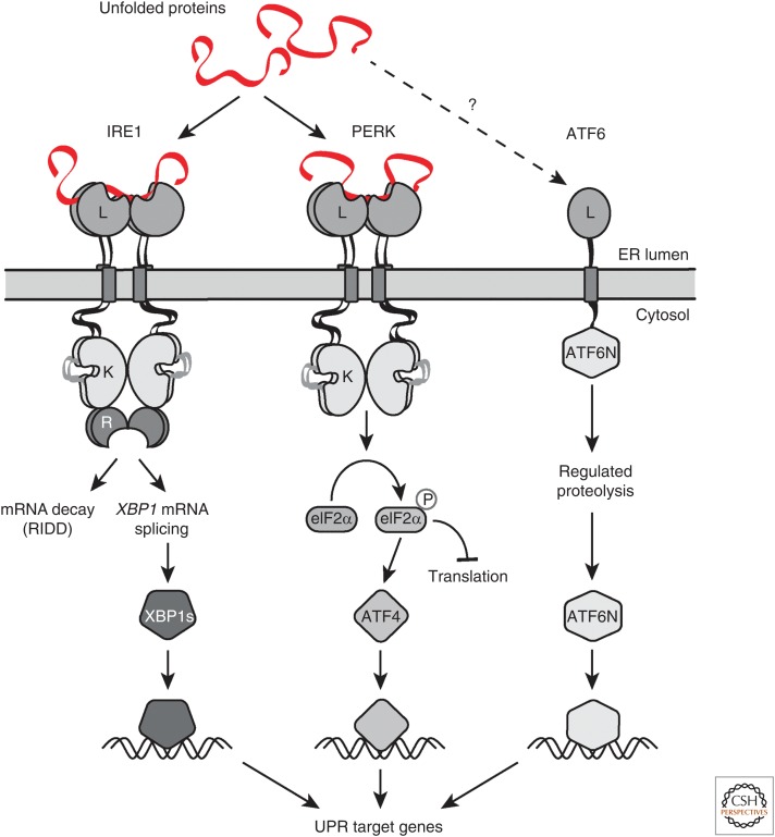
Most of the secreted and plasma membrane proteins are synthesized on membrane-bound ribosomes on the endoplasmic reticulum (ER). They require engagement of ER-resident chaperones and foldases that assist in their folding and maturation. Since protein homeostasis in the ER is crucial for cellular function, the protein-folding status in the organelle's lumen is continually surveyed by a network of signaling pathways, collectively called the unfolded protein response (UPR). Protein-folding imbalances, or "ER stress," are detected by highly conserved sensors that adjust the ER's protein-folding capacity according to the physiological needs of the cell. We review recent developments in the field that have provided new insights into the ER stress-sensing mechanisms used by UPR sensors and the mechanisms by which they integrate various cellular inputs to adjust the folding capacity of the organelle to accommodate to fluctuations in ER protein-folding demands.


Figures

Figure 1.
Figure 1.
Three branches of the unfolded protein response (UPR). Three endoplasmic reticulum (ER) stress sensors, IRE1, PERK, and ATF6, monitor the protein-folding conditions in the ER lumen. Each pathway uses a unique mechanism of signal transduction that results in the activation of specialized transcription factors that drive transcription of target genes that alleviate ER stress. The IRE1 and PERK branches also reduce the ER-folding load by impeding further client protein load in the ER either by degrading ER-targeted messenger RNAs (mRNAs) or by negatively regulating translation, respectively.
Figure 2.
Figure 2.
IRE1's endoplasmic reticulum (ER) protein-folding stress-sensing mechanism. In steady-state conditions, the ER-resident chaperone BiP binds IRE1's sensor domain and shifts IRE1 to a monomeric inactive state, buffering IRE1's activity. During ER stress, BiP is titrated to unfolded proteins accumulating in the ER, relieving its buffering effect on IRE1. Simultaneously, unfolded proteins are directly recognized by IRE1's sensor lumenal domain. These unfolded proteins serve as activating ligands that drive IRE1 oligomerization and activation.
Figure 3.
Figure 3.
Comparison between the crystal structures of the sensor domains of IRE1 and PERK. Crystal structures of the core lumenal domains (cLDs) of yeast (A), and human IRE1 (B), as well as that of the human PERK cLD (C) reveal that key structural elements forming the dimerization interface (endoplasmic reticulum [ER]-lumenal interface 1, IF1L; indicated by dashed lines) are highly conserved among yeast and mammalian IRE1 as well as in the mammalian PERK cLD. By contrast, the α-helix turn forming oligomerization interface 2 (IF2L, indicated by the black arrow) in the yeast IRE1 cLD is not conserved in the mammalian ER stress sensors. The structures are shown in colored ribbon diagram representations on the left and as surface representations in grey on the right. The major histocompatibility complex (MHC)-like groove in the yeast IRE1 cLD structure is shaded in red. The αB helix found only in human IRE1α cLD is indicated with an arrow. The distance between the helices surrounding the groove is depicted with double-pointed black arrows.
Figure 4.
Figure 4.
Apo-human IRE1 lumenal domain (LD) dimers are found in equilibrium between closed and open conformations (step 1). Upon endoplasmic reticulum (ER) stress, unfolded proteins accumulating in the ER lumen bind the IRE1 LD, stabilizing the sensor domain in its open conformation. Peptide binding also induces a conformational change in the αB helix and the neighboring structural elements (steps 2 and 3), that facilitate activation by allowing the formation of an IF2L-like interface in the protein's LD (step 4). When protein-folding homeostasis is achieved, the dynamic IRE1 LD oligomers re-adopt their inactive conformation (step 5).

References

    1. Acosta-Alvear D, Zhou Y, Blais A, Tsikitis M, Lents NH, Arias C, Lennon CJ, Kluger Y, Dynlacht BD. 2007. XBP1 controls diverse cell type- and condition-specific transcriptional regulatory networks. Mol Cell 27: 53–66. 10.1016/j.molcel.2007.06.011 - DOI - PubMed
    1. Adachi Y, Yamamoto K, Okada T, Yoshida H, Harada A, Mori K. 2008. ATF6 is a transcription factor specializing in the regulation of quality control proteins in the endoplasmic reticulum. Cell Struct Funct 33: 75–89. 10.1247/csf.07044 - DOI - PubMed
    1. Adamson B, Norman TM, Jost M, Cho MY, Nunez JK, Chen Y, Villalta JE, Gilbert LA, Horlbeck MA, Hein MY, et al. 2016. A multiplexed single-cell CRISPR screening platform enables systematic dissection of the unfolded protein response. Cell 167: 1867–1882.e21. 10.1016/j.cell.2016.11.048 - DOI - PMC - PubMed
    1. Amin-Wetzel N, Saunders RA, Kamphuis MJ, Rato C, Preissler S, Harding HP, Ron D. 2017. A J-protein cochaperone recruits BiP to monomerize IRE1 and repress the unfolded protein response. Cell 171: 1625–1637.e13. 10.1016/j.cell.2017.10.040 - DOI - PMC - PubMed
    1. Anelli T, Sitia R. 2008. Protein quality control in the early secretory pathway. EMBO J 27: 315–327. 10.1038/sj.emboj.7601974 - DOI - PMC - PubMed