Review
. 2019 Aug 1;11(8):a033928.
doi: 10.1101/cshperspect.a033928.

Chaperoning Endoplasmic Reticulum-Associated Degradation (ERAD) and Protein Conformational Diseases

Affiliations
Review

Chaperoning Endoplasmic Reticulum-Associated Degradation (ERAD) and Protein Conformational Diseases

Patrick G Needham et al. Cold Spring Harb Perspect Biol. .

Abstract

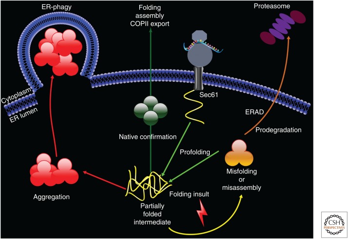
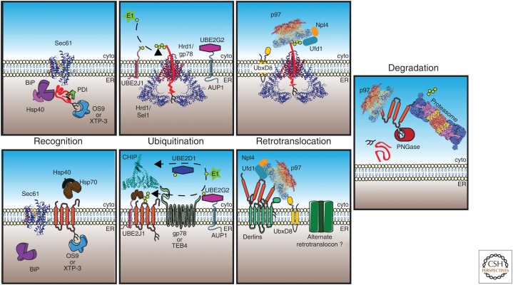
Misfolded proteins compromise cellular homeostasis. This is especially problematic in the endoplasmic reticulum (ER), which is a high-capacity protein-folding compartment and whose function requires stringent protein quality-control systems. Multiprotein complexes in the ER are able to identify, remove, ubiquitinate, and deliver misfolded proteins to the 26S proteasome for degradation in the cytosol, and these events are collectively termed ER-associated degradation, or ERAD. Several steps in the ERAD pathway are facilitated by molecular chaperone networks, and the importance of ERAD is highlighted by the fact that this pathway is linked to numerous protein conformational diseases. In this review, we discuss the factors that constitute the ERAD machinery and detail how each step in the pathway occurs. We then highlight the underlying pathophysiology of protein conformational diseases associated with ERAD.


Figures

Figure 1.
Figure 1.
Nascent polypeptide biogenesis in the endoplasmic reticulum (ER). After docking at the ER membrane on the Sec61 translocation channel, the ribosome cotranslationally translocates a polypeptide (in yellow) into the ER lumen. Chaperones, including Hsp70s, Hsp40s, NEFs, lectins, disulfide isomerases, and peptidyl proline isomerase, act as “profolding” factors (see text for details). If the polypeptide acquires its native conformation and—as shown in one example, oligomerizes (in green)—it traffics from the ER in COPII vesicles. Genetic mutations, errors in transcription or translation, defects in the acquisition of posttranslational modifications, and/or ER stress can result in misfolding or a misassembled intermediate (in orange). If the substrate is unable to fold, “prodegradation” chaperones facilitate ER-associated degradation (ERAD). In contrast, partially folded intermediates can aggregate (in red) and are instead cleared by ER-phagy. One route for this pathway leads to the formation of an “omegasome,” as depicted (Simonsen and Stenmark 2008). For simplicity, only a soluble ER lumenal protein is presented.
Figure 2.
Figure 2.
Endoplasmic reticulum-associated degradation (ERAD) of lumenal and integral membrane substrates. (Top) During translocation through Sec61 (RCSB PDB 3JC2), a soluble ERAD substrate (in red) encounters BiP, which aids in polypeptide folding and translocation across the membrane. BiP activity is enhanced by ER lumenal Hsp40s. The polypeptide can be modified by the formation of disulfide bonds by protein disulfide isomerases (PDIs) and by addition of N-linked glycans, which are bound by lectins such as OS9 or XTP-3 (in mammals). If the polypeptide fails to fold, it is transferred to Hrd1. The E3 ligase/RING domain of Hrd1 is located in the cytoplasm, so lumenal substrates must be partially retrotranslocated for ubiquitin conjugation, which occurs via an enzymatic cascade that includes an E1 (ubiquitin-activating enzyme), E2 (ubiquitin-conjugating enzyme such as UBE2G2/AUP1 or UBE2J1), and E3 (ubiquitin ligase such as Hrd1 or gp78), which build a polyubiquitin chain (lime hexagons) on the substrate. AUP1 tethers the E2 to the ER membrane and functions analogously to Cue1 in yeast. Following ubiquitination, the substrate is bound by the p97 (RCSB PDB 5C1A) complex, which is recruited to the membrane by UbxD8 and interacts with the substrate via ubiquitin-binding partners Ufd1 and Npl1. (Bottom) Integral membrane ERAD substrates (in orange) may also interact with BiP, but also have access to cytoplasmic chaperones such as Hsp70-Hsp40 that recognize folding lesions. Cytoplasmic lysines are then polyubiquitinated by E3 ligases such as TEB4, gp78, and/or CHIP (cyan, RCSB PDB 2C2L) in mammals. The p97–Ufd1–Npl1 heterotrimer is recruited by UbxD8 and, in some cases, Derlin family members, which facilitate retrotranslocation. The nature of the retrotranslocon channel (green) for membrane proteins is not established. Finally, the retrotranslocated protein can be deglycosylated by PNGase, followed by delivery to the 26S proteasome (RCSB PDB 4CR2), deubiquitinated by components in the 19S particle (purple/blue), and proteolyzed in the 20S core particle (yellow/salmon).

References

    1. Accili D, Kadowaki T, Kadowaki H, Mosthaf L, Ullrich A, Taylor SI. 1992. Immunoglobulin heavy chain-binding protein binds to misfolded mutant insulin receptors with mutations in the extracellular domain. J Biol Chem 267: 586–590. - PubMed
    1. Aebi M, Bernasconi R, Clerc S, Molinari M. 2010. N-glycan structures: Recognition and processing in the ER. Trends Biochem Sci 35: 74–82. 10.1016/j.tibs.2009.10.001 - DOI - PubMed
    1. Allen S, Abuzenadah AM, Hinks J, Blagg JL, Gursel T, Ingerslev J, Goodeve AC, Peake IR, Daly ME. 2000. A novel von Willebrand disease–causing mutation (Arg273Trp) in the von Willebrand factor propeptide that results in defective multimerization and secretion. Blood 96: 560–568. - PubMed
    1. Allen S, Goodeve AC, Peake IR, Daly ME. 2001. Endoplasmic reticulum retention and prolonged association of a von Willebrand's disease-causing von Willebrand factor variant with ERp57 and calnexin. Biochem Biophys Res Commun 280: 448–453. 10.1006/bbrc.2000.4139 - DOI - PubMed
    1. Allen JR, Nguyen LX, Sargent KEG, Lipson KL, Hackett A, Urano F. 2004. High ER stress in β-cells stimulates intracellular degradation of misfolded insulin. Biochem Biophys Res Commun 324: 166–170. 10.1016/j.bbrc.2004.09.035 - DOI - PubMed

Publication types

LinkOut - more resources