. 2019 Apr 15;25(8):2644-2655.
doi: 10.1158/1078-0432.CCR-18-1401. Epub 2019 Jan 22.

Role of Tumor-Associated Macrophages in the Clinical Course of Pancreatic Neuroendocrine Tumors (PanNETs)

Affiliations

Role of Tumor-Associated Macrophages in the Clinical Course of Pancreatic Neuroendocrine Tumors (PanNETs)

Lei Cai et al. Clin Cancer Res. .

Abstract

Purpose: This study evaluated the potential role of immune cells and molecules in the pathogenesis and clinical course of pancreatic neuroendocrine tumors (PanNET).

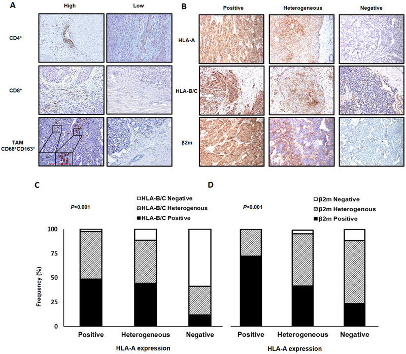
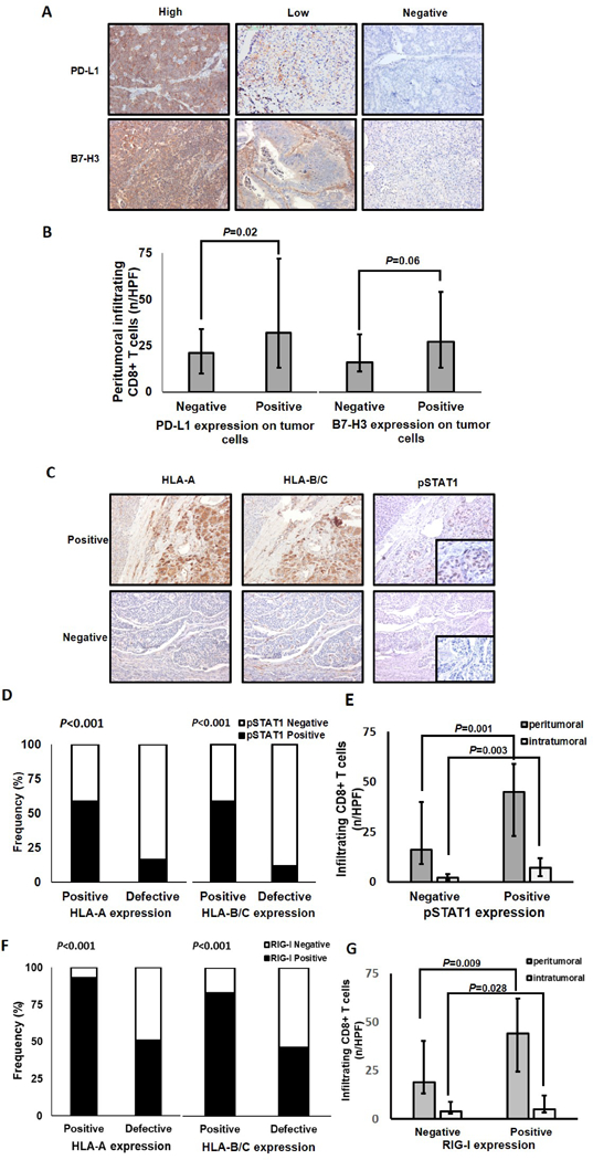
Experimental design: Surgically resected PanNETs (N = 104) were immunohistochemically analyzed for Ki67 index, mitotic rate, macrophage, CD4+ cells, and CD8+ T-cell infiltration, as well as HLA class I, PD-L1, and B7-H3 expression. Results were correlated with clinicopathologic characteristics as well as with disease-free (DFS) and disease-specific (DSS) survival.

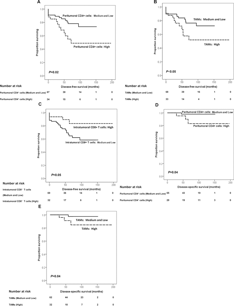
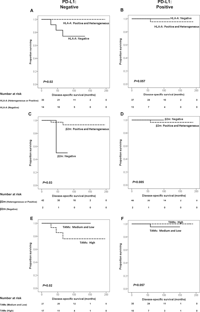
Results: The median age of the 57 WHO grade 1 and 47 WHO grade 2 patients was 55 years. High intratumoral CD8+ T-cell infiltration correlated with prolonged DFS (P = 0.05), especially when the number of tumor-associated macrophages (TAM) was low. In contrast, high peritumoral CD4+ cell and TAM infiltration was associated with a worse DFS and DSS. PD-L1 and B7-H3 were expressed in 53% and 78% PanNETs, respectively. HLA class I expression was defective in about 70% PanNETs. HLA-A expression correlated with favorable DSS in PD-L1-negative tumors (P = 0.02). TAM infiltration (P = 0.02), WHO grade (P = 0.04), T stage (P = 0.01), and lymph node positivity (P = 0.04) were independent predictors of DFS. TAM infiltration (P = 0.026) and T stage (P = 0.012) continued to be predictors of DFS in WHO grade 1 PanNET patients. TAM infiltration was the sole independent predictor of DSS for WHO grade 1 and 2 patients (P = 0.02). Therefore, this biomarker may contribute to identifying WHO grade 1 patients with poor prognosis.

Conclusions: TAM infiltration appears to be the most informative prognostic biomarker in PanNET. It may represent a useful immunotherapeutic target in patients with PanNET.


Conflict of interest statement

Conflicts of Interest: The authors declare no potential conflicts of interest.

Figures

Figure 1.
Figure 1.
Immune cell infiltration and HLA class I expression in PanNET tumors. A, PanNET tissue sections were IHC stained with rabbit anti-CD4, mouse anti-CD8, mouse anti-CD68, and rabbit anti-CD163 mAbs. Different levels of CD4+ cell, CD8+ T cell, and TAM (CD68+CD163+) infiltration (X200 original magnification, insets X800 original magnification) are shown. B, PanNET tissue sections were IHC stained with mouse anti-HLA-A mAb HCA2, anti-HLA-B/C mAb HC-10, and anti-β2m mAb NAMB-1. Representative patterns of positive, heterogeneous, and negative HLA-A, HLA-B/C and β2m staining (X200 original magnification) are shown. C, HLA-A expression is significantly associated with HLA-B/C expression (P<0.001). D, HLA-A expression is significantly associateted with β2m expression (P<0.001). P values derived from Fisher exact test.
Figure 2.
Figure 2.
Association of PD-L1, B7-H3, phospho-STAT1, and RIG-I expression with the extent of peritumoral CD8+T-cell infiltration and with HLA class I expression in PanNETs. A, PanNET tissue sections were IHC stained with rabbit anti-PD-L1 mAb and goat anti-B7-H3 pAb. Representative patterns of highly positive embrane/cytoplasmic, scattered positive membrane/cytoplasmic and negative PD-L1 staining (X200 original magnification) are shown. Representative patterns of highly positive membrane/cytoplasmic, low membrane/cytoplasmic and negative B7-H3 staining (X200 original magnification) are shown. B, Peritumoral CD8+ T-cell number is significantly associated with PD-L1 expression (P=0.02), but not with cytoplasmic and/or membranous B7-H3 expression (P=0.06). C, PanNET tissue sections were IHC stained with mouse anti-HLA-A mAb HCA2, mouse anti-HLA-B/C mAb HC-10, rabbit anti-phosphoStat1 (pSTAT1) mAb, and rabbit anti-RIG-I pAb. The positive HLA-A and HLA-B/C expression in PanNETs with positive pSTAT1 expression and the low/negative HLA-A and HLA-B/C expression in PanNETs with negative phosphoStat1 expression (X200 original magnification, insets X400 original magnification) are shown. D, Positive HLA-A and HLA-B/C expression is significantly associated with positive pSTAT1 expression (P<0.001). E, Peritumoral and intratumoral CD8+ T-cell number is significantly associated with pSTAT1 expression (P=0.001 and P=0.003). F, HLA-A and HLA-B/C expression is significantly associated with RIG-I expression (P<0.001). G, Peritumoral and intratumoral CD8+ T-cell number is significantly associated with positive RIG-I expression (P=0.009 and P=0.028). Bars, median values. Error bars, interquartile range. P values derived from Mann–Whitney U tests.
Figure 3.
Figure 3.
Association of the extent of tumor infiltration by immune cells with DFS and DSS in patients with PanNET. DFS based on peritumoral CD4+ cell, TAM, and intratumoral CD8+ T-cell number is shown. Prognosis is depicted with Kaplan–Meier curves: (A) for peritumoral CD4+ cell number: high versus medium and low (mean survival: high: 113 vs. medium and low: 123 months, P=0.02), (B) for TAM number: high versus medium and low (mean survival: high: 113 versus medium and low: 121 months, P<0.05); and (C) for intratumoral CD8+ T-cell number: high versus medium and low (mean survival: high: 170 vs. medium and low: 104 months, P=0.05). Disease-specific survival based on peritumoral CD4+ cells and TAM number is shown. Prognosis is depicted with Kaplan–Meier curves: (D) for peritumoral CD4+ cell number: high versus medium and low (mean survival: high: 150 vs. medium and low: 173 months, P=0.04), and (E) for TAM number: high versus medium and low (mean survival: high: 149 vs. medium and low: 175 months, P =0.04). P values derived from log-rank tests.
Figure 4.
Figure 4.
Impact of PD-L1 expression on the association of HLA-A and β2m expression by PanNET cells and of TAM number with DSS in patients with PanNET. A and B, Prognosis is depicted with Kaplan-Meier curves for HLA-A expression (positive and heterogeneous vs. negative) in PD-L1-negative (P=0.02) and positive (P=0.057) group. C and D, Prognosis is depicted with Kaplan-Meier curves for β2m expression (positive and heterogeneous vs. negative) in PD-L1–negative (P=0.03) and positive (P=0.085) group. E and F, Prognosis is depicted with Kaplan-Meier curves for TAM number (high vs. medium and low) in PD-L1-negative (P=0.02) and positive (P=0.057) group. P values derived from logrank tests.

References

    1. Halfdanarson TR, Rubin J, Farnell MB, Grant CS, Petersen GM. Pancreatic endocrine neoplasms: epidemiology and prognosis of pancreatic endocrine tumors. Endocr Relat Cancer 2008;15(2):409–27 doi 10.1677/ERC-07-0221. - DOI - PMC - PubMed
    1. Metz DC, Jensen RT. Gastrointestinal neuroendocrine tumors: pancreatic endocrine tumors. Gastroenterology 2008;135(5):1469–92 doi 10.1053/j.gastro.2008.05.047. - DOI - PMC - PubMed
    1. Cloyd JM, Poultsides GA. Non-functional neuroendocrine tumors of the pancreas: Advances in diagnosis and management. World J Gastroenterol 2015;21(32):9512–25 doi 10.3748/wjg.v21.i32.9512. - DOI - PMC - PubMed
    1. Dromain C, Deandreis D, Scoazec JY, Goere D, Ducreux M, Baudin E, et al. Imaging of neuroendocrine tumors of the pancreas. Diagn Interv Imaging 2016;97(12):1241–57 doi 10.1016/j.diii.2016.07.012. - DOI - PubMed
    1. Klimstra DS, Modlin IR, Coppola D, Lloyd RV, Suster S. The pathologic classification of neuroendocrine tumors: a review of nomenclature, grading, and staging systems. Pancreas 2010;39(6):707–12 doi 10.1097/MPA.0b013e3181ec124e. - DOI - PubMed

Publication types

MeSH terms