Comparative Study
. 2019 Mar 25;87(4):e00758-18.
doi: 10.1128/IAI.00758-18. Print 2019 Apr.

Atypical Hemolytic Listeria innocua Isolates Are Virulent, albeit Less than Listeria monocytogenes

Affiliations
Comparative Study

Atypical Hemolytic Listeria innocua Isolates Are Virulent, albeit Less than Listeria monocytogenes

Alexandra Moura et al. Infect Immun. .

Abstract

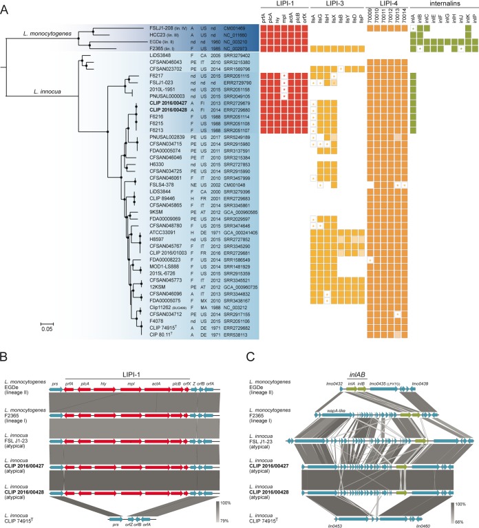
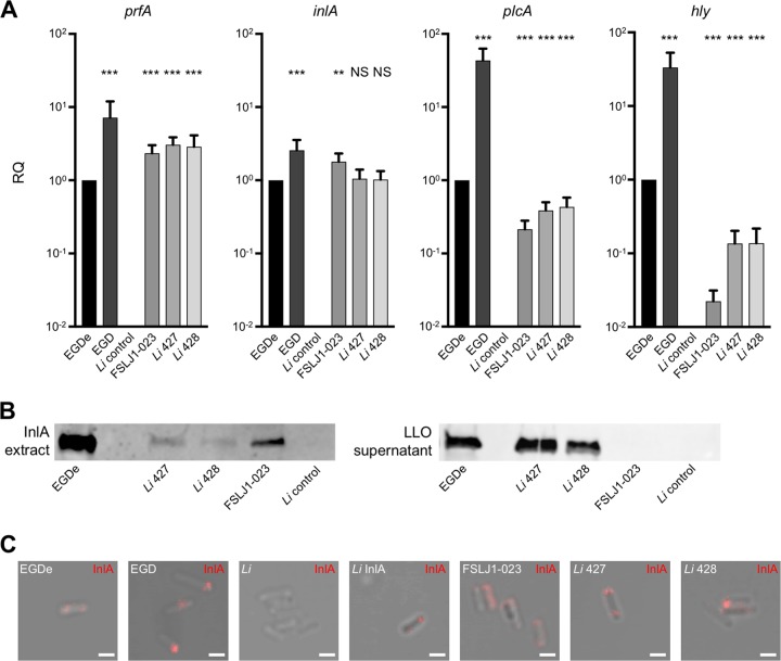
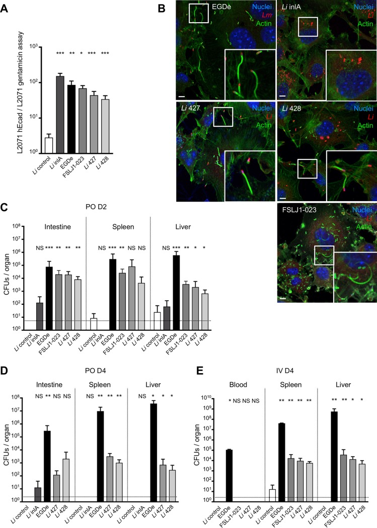
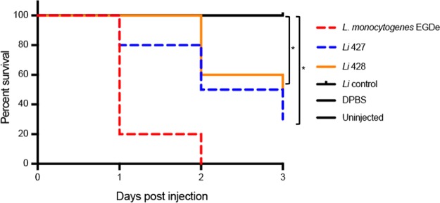
Listeria innocua is considered a nonpathogenic Listeria species. Natural atypical hemolytic L. innocua isolates have been reported but have not been characterized in detail. Here, we report the genomic and functional characterization of representative isolates from the two known natural hemolytic L. innocua clades. Whole-genome sequencing confirmed the presence of Listeria pathogenicity islands (LIPI) characteristic of Listeria monocytogenes species. Functional assays showed that LIPI-1 and inlA genes are transcribed, and the corresponding gene products are expressed and functional. Using in vitro and in vivo assays, we show that atypical hemolytic L. innocua is virulent, can actively cross the intestinal epithelium, and spreads systemically to the liver and spleen, albeit to a lesser degree than the reference L. monocytogenes EGDe strain. Although human exposure to hemolytic L. innocua is likely rare, these findings are important for food safety and public health. The presence of virulence traits in some L. innocua clades supports the existence of a common virulent ancestor of L. monocytogenes and L. innocua.

Keywords: L. innocua; LIPI; inlA; listeriosis; virulence.


Figures

FIG 1
FIG 1
Phylogenetic and comparative genomic analyses. (A) Rooted maximum likelihood phylogeny of 42 L. innocua genomes based on 642,408 core genome SNPs. Representative genomes of the four L. monocytogenes lineages were used as the outgroup. Circles represent bootstrap branch support values higher than 90% based on 1,000 replicates. Information on the source (A, animal; F, food; H, human; PE, production environment; NE, natural environment; nd, unknown), country, and year of isolation, as well as the NCBI/EMBL/DDBJ accession numbers, is provided in the columns. Colored boxes represent the presence of the different genetic traits. Light-colored boxes represent genes interrupted by end of contigs. Stars represent truncated genes due to the presence of internal stop codons. (B and C) Organization of the LIPI-1 and inlAB loci and their flanking regions. Arrows denote the orientation of genes. Gray blocks denote BLASTN similarities between sequences.
FIG 2
FIG 2
Expression of LIPI-1 and inlA genes in atypical L. innocua. (A) qRT-PCR quantification of prfA, inlA, plcA, and hly transcripts produced in BHI broth at 37°C by L. monocytogenes (EGDe and EGD), L. innocua CLIP 74915T (Li control), and the atypical L. innocua CLIP 2016/00427 (Li 427) and CLIP 2016/00428 (Li 428). Each strain was tested at least three times using independent precultures. gyrB was used as a stable reference gene for normalization. Results are shown as fold change of prfA, inlA, plcA, and hly expression relative to that of EGDe (RQ, relative quantities). A Mann-Whitney test was performed; strains were compared to EGDe (NS, not significant; **, P < 0.01; ***, P < 0.001). (B) InlA and LLO Western blotting of 30 μg proteins of the exponential-phase culture supernatants of each isolate. Immunoblots were performed at least two times. To avoid saturation of signal, strain EGD was not included in this analysis. (C) Cell surface exposure of InlA. Immunofluorescence analysis was performed with mouse monoclonal anti-InlA antibodies revealed with an anti-IgG coupled to Alexa Fluor 555. Scale, 5 μm.
FIG 3
FIG 3
In vitro and in vivo functionality of LIPI-1 and inlA in atypical L. innocua. (A) Gentamicin assay of L. monocytogenes EGDe, L. innocua CLIP 74915T (Li control), L. innocua pAD-inlA mutant (Li inlA), and atypical L. innocua CLIP 2016/00427 (Li 427) and CLIP 2016/00428 (Li 428) in mouse fibroblast L2071 and L2071 hEcad. CFU of gentamicin-resistant bacteria were enumerated. Invasion assays were performed in triplicates and represent at least three independent experiments for each condition tested. Ratio represents CFU in L2071 hEcad divided by CFU in L2071 for each strain. (B) Abilities of L. monocytogenes EGDe, L. innocua pAD-inlA mutant (Li inlA), and atypical L. innocua CLIP 2016/00427 (Li 427) and CLIP 2016/00428 (Li 428) to polymerize host actin were compared after invasion assay on L2071 hEcad cells. Bacteria were detected with anti-L. monocytogenes or anti-L. innocua (red), actin with phalloidin (green), and nuclei with Hoechst (blue). Scale, 5 μm. (C) In vivo assay in KI E16P female mice (n = 6 mice for each condition, n =10 for Li inlA and EGDe) infected orally with 5 × 10⁹ CFU and dissected 48 h after infection. PO, per os. (D) In vivo assay in KI E16P female mice (n = 6 mice for each condition) infected orally with 5 × 10⁹ CFU and dissected 96 h after infection. (E) In vivo assay in KI E16P female mice (n = 6 mice for each condition) infected intravenously (IV) with 5 × 10⁴ CFU and dissected 96 h after infection. For panels A, C, D, and E, a Mann-Whitney test was performed; strains were compared to L. innocua CLIP 74915T (Li control) (NS, not significant; *, P < 0.05; **, P < 0.01; ***, P < 0.001). Values represent means ± standard deviations.
FIG 4
FIG 4
Infection assays in zebrafish. Survival rates of zebrafish embryos after inoculation with L. monocytogenes EGDe, L. innocua (Li control), and atypical L. innocua CLIP 2016/00427 (Li 427) and CLIP 2016/00428 (Li 428) bacterial suspensions in DPBS (n = 30; *, P < 0.05 for comparisons relative to the L. innocua control strain).

References

    1. Orsi RH, Wiedmann M. 2016. Characteristics and distribution of Listeria spp., including Listeria species newly described since 2009. Appl Microbiol Biotechnol 100:5273–5287. doi:10.1007/s00253-016-7552-2. - DOI - PMC - PubMed
    1. Seeliger HP. 1981. Nonpathogenic listeriae: L. innocua sp. n. (Seeliger et Schoofs, 1977) (author’s transl). Zentralbl Bakteriol Mikrobiol Hyg A 249:487–493. doi:10.1016/S0174-3031(81)80108-4. - DOI - PubMed
    1. Charlier C, Perrodeau É, Leclercq A, Cazenave B, Pilmis B, Henry B, Lopes A, Maury MM, Moura A, Goffinet F, Dieye HB, Thouvenot P, Ungeheuer MN, Tourdjman M, Goulet V, de Valk H, Lortholary O, Ravaud P, Lecuit M, MONALISA Study Group. 2017. Clinical features and prognostic factors of listeriosis: the MONALISA national prospective cohort study. Lancet Infect Dis 17:510–519. doi:10.1016/S1473-3099(16)30521-7. - DOI - PubMed
    1. Perrin M, Bemer M, Delamare C. 2003. Fatal case of Listeria innocua bacteremia. J Clin Microbiol 41:5308–5309. doi:10.1128/JCM.41.11.5308-5309.2003. - DOI - PMC - PubMed
    1. Favaro M, Sarmati L, Sancesario G, Fontana C. 2014. First case of Listeria innocua meningitis in a patient on steroids and eternecept. JMM Case Rep 1:1–5. doi:10.1099/jmmcr.0.003103. - DOI

Publication types

MeSH terms

Substances