. 2019 Jan 22;10(1):236.
doi: 10.1038/s41467-018-08264-w.

Improved TMC1 gene therapy restores hearing and balance in mice with genetic inner ear disorders

Affiliations

Improved TMC1 gene therapy restores hearing and balance in mice with genetic inner ear disorders

Carl A Nist-Lund et al. Nat Commun. .

Erratum in

Abstract

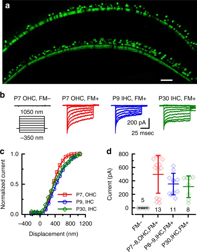
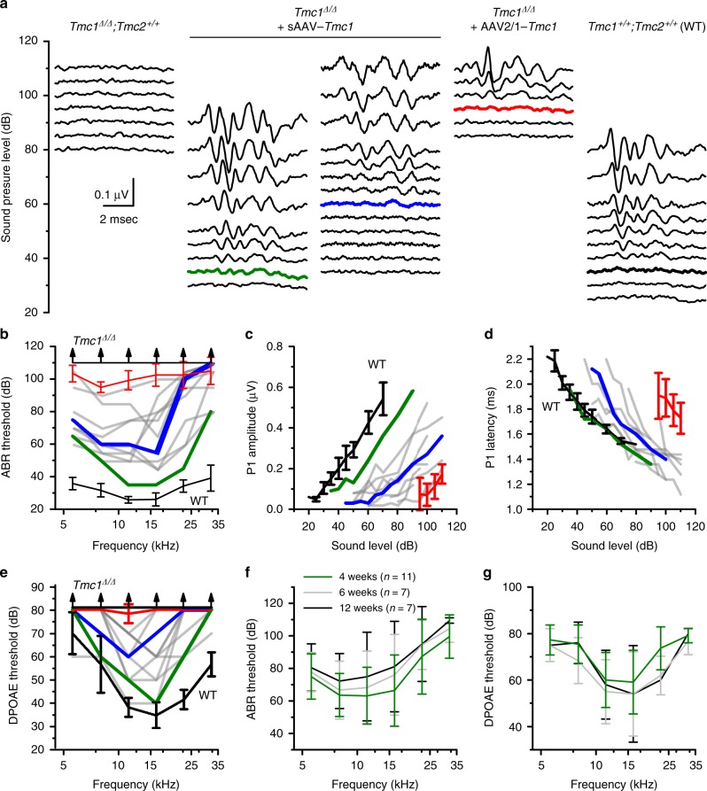
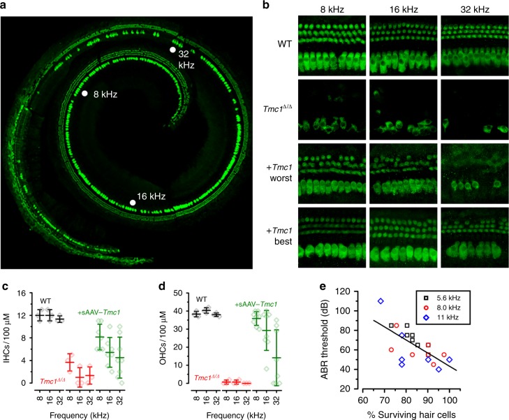
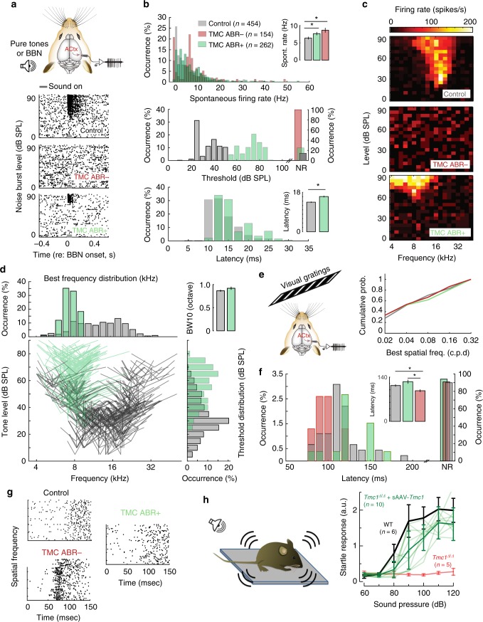
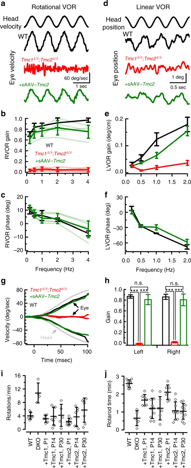
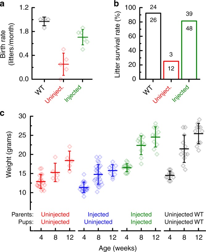
Fifty percent of inner ear disorders are caused by genetic mutations. To develop treatments for genetic inner ear disorders, we designed gene replacement therapies using synthetic adeno-associated viral vectors to deliver the coding sequence for Transmembrane Channel-Like (Tmc) 1 or 2 into sensory hair cells of mice with hearing and balance deficits due to mutations in Tmc1 and closely related Tmc2. Here we report restoration of function in inner and outer hair cells, enhanced hair cell survival, restoration of cochlear and vestibular function, restoration of neural responses in auditory cortex and recovery of behavioral responses to auditory and vestibular stimulation. Secondarily, we find that inner ear Tmc gene therapy restores breeding efficiency, litter survival and normal growth rates in mouse models of genetic inner ear dysfunction. Although challenges remain, the data suggest that Tmc gene therapy may be well suited for further development and perhaps translation to clinical application.


Conflict of interest statement

J.R.H. is a consultant to several companies focused on development of inner ear therapeutics. J.R.H. and G.S.G. hold patents on Tmc1 gene therapy which have not been licensed. The remaining authors declare no competing interests.

Figures

Fig. 1
Fig. 1
sAAV-Tmc1 restores sensory transduction in Tmc1∆/∆;Tmc2∆/∆ IHCs and OHCs. a Confocal images of two apical sections excised from P7 Tmc1∆/∆;Tmc2∆/∆ mouse cochleas after injection of sAAV-Tmc1 through the RWM at P1. The tissue was cultured for 7 days and perfused with 5 µM FM1-43 for 10 s followed by 3 full bath exchanges to washout excess dye. The tissue was then mounted and imaged for FM1-43 uptake (green) in IHCs and OHCs. Scale bar = 25 µm. b Representative families of sensory transduction currents evoked by mechanical displacement of hair bundles (protocol shown below) recorded from P7 apical OHCs of Tmc1∆/∆;Tmc2∆/∆ mice injected with sAAV-Tmc1 that were FM1-43-negative (left) or FM1-43-positive (second from left). FM1-43-positive currents were also recorded from P9 IHCs (second from right) and P30 IHCs (right) of Tmc1∆/∆;Tmc2∆/∆ mice injected with sAAV-Tmc1. c Stimulus-response curves from the currents shown in b. d Mean ± S.D. peak sensory transduction current amplitudes from apical FM1-43-negative and FM1-43-positive IHCs and OHCs from Tmc1∆/∆;Tmc2∆/∆ mice injected with sAAV-Tmc1 at time points as indicated. n = number of hair cells
Fig. 2
Fig. 2
sAAV-Tmc1 restores ABRs and DPOAEs in Tmc1∆/∆ mice. a Families of ABR waveforms recorded at P28-P30 from uninjected Tmc1∆/∆ mouse (left) and from Tmc1∆/∆ mice injected with sAAV-Tmc1 or AAV2/1-Tmc1 and one WT control, as indicated above. ABRs were recorded using 11.3-kHz tone bursts at sound pressure levels increasing by 5-dB until peak amplitudes reached 0.55 μV. Thresholds determined by the presence of Peak 1 and is indicated by colored traces. Scale bar applies to all families. b ABR thresholds plotted as a function of stimulus frequency for eleven Tmc1∆/∆ mice injected with sAAV-Tmc1 tested at P28-P30 (gray traces). Best (green), and median (blue) recovery are indicated. Black lines show mean ± S.D. (n= 6 mice) for WT and Tmc1∆/∆ uninjected mice as indicated. Red line shows mean ± S.D. (n= 6 mice) for Tmc1∆/∆ injected with AAV2/1-Tmc1. c Peak 1 amplitudes measured from 11.3-kHz ABR waveforms, as shown in a, for eleven Tmc1∆/∆ mice injected with sAAV-Tmc1. Colors correspond to conditions indicated in b. d Peak 1 latencies measured from 11.3-kHz ABR waveforms, as shown in a for eleven Tmc1∆/∆ mice injected with sAAV-Tmc1. Colors correspond to conditions indicated in b. e DPOAE thresholds plotted as a function of stimulus frequency for eleven Tmc1∆/∆ mice injected with sAAV-Tmc1 tested at P28-P30. Colors correspond to conditions indicated in b. f Mean ± S.D. (n = number of mice) ABR thresholds plotted as a function of stimulus frequency for Tmc1∆/∆ mice injected with sAAV-Tmc1 at P1 tested at 4, 6, and 12 weeks. g Mean ± S.D. DPOAE thresholds plotted as a function of stimulus frequency for the same Tmc1∆/∆ mice shown in panel G injected with sAAV-Tmc1
Fig. 3
Fig. 3
sAAV-Tmc1 promotes survival of IHCs and OHCs in Tmc1∆/∆ mice. a Composite of five images from a whole cochlea harvested at 12 weeks old from a Tmc1∆/∆ mouse injected with sAAV-Tmc1 on P1 reconstructed from microdissected tissue. The tissue was stained for Myo7a (green). b Tonotopically mapped cochlear sections (image width = 100 µm) at 8, 16, and 32 kHz regions for WT (top), Tmc1∆/∆ (second), and Tmc1∆/∆-injected with sAAV-Tmc1 on P1 with the worst (third) and best (bottom) ABR performances as indicated in Fig. 2. c, d The number of surviving IHCs (c) and OHCs (d) in sections indicated in b from 3 WT  mice, 3 uninjected Tmc1∆/∆, and eight Tmc1∆/∆ mice injected with sAAV-Tmc1 on P1 showing mean ± S.D. Number of surviving IHCs in the 8 and 16 kHz was significantly greater in injected ears than in uninjected Tmc1∆/∆ ears at (t-test: p = 0.015, 0.03, 0.19, respectively). Number of surviving OHCs in the 8 and 16 kHz was significantly greater in injected ears than in uninjected Tmc1∆/∆ ears at (t-test: p = 1E−7, 0.002, 0.16, respectively). e The percentage of surviving hair cells at 12 weeks of age was correlated with ABR thresholds measured between 5.6 and 11 kHz from sAAV-Tmc1-injected Tmc1∆/∆ mice. The data were fit with a linear regression with r = −0.67 and p = 6.2 × 10-4 (n = 7 mice; 21 measurements)
Fig. 4
Fig. 4
sAAV-Tmc1 restores neuronal responses in auditory cortex of Tmc1∆/∆ mice. a (Top) Schematic of multi-unit (MU) activity recordings in the auditory cortex. (Below) Representative raster plots from each group. Data were recorded from six C57B/L6 wild-type control mice and eleven awake head-fixed Tmc1∆/∆ mice injected with sAAV-Tmc1. Tmc1∆/∆-injected mice were divided into two groups, those with significant improvement in ABR thresholds (ABR+: thresholds ≤70 dB at any frequency; n = 7 mice) and mice with little improvement in thresholds (ABR−: thresholds ≥75 dB at all frequencies; n = 4 mice). Sound stimulus duration indicated as gray bar. BBN: broad band noise, ACtx: auditory cortex. b Quantification of spontaneous activity for WT control mice and Tmc1∆/∆-injected mice (top). Quantification of both threshold (middle) and latency (bottom) resulting from broad band noise (BBN) stimuli presentation at different sound levels. c Representative frequency response areas after presentation of 361 frequency-level sound combinations. d Distribution of population tuning properties for responsive units Illustrating the best frequency (tip) and bandwidth measured 10 dB above threshold (side lines). Distribution of pure tone thresholds (bottom, right) and best frequencies (top, left) for responsive units. (Top, right) Bandwidth tuning responses for the stimulus presentation in (C). e (Top left) Schematic of recordings from auditory cortex during presentation of a visual stimulus consisting of gratings with changing spatial frequency. (Right) Preferred spatial frequency among ABR+, ABR−, and control mice. f Quantification of visually evoked onset latency in ABR− mice, ABR+ and control mice. g Representative raster plots from neurons recorded from the paradigm in f. h Startle response amplitudes normalized to mouse weight measured at P28-P30 and plotted as a function of sound intensity and as mean ± S.D. for 6 control C57BL/6 mice (black), ten individual Tmc1∆/∆ mice injected with sAAV-Tmc1 (light green, mean ± S.D. in bold green), and five uninjected Tmc1∆/∆ mice (red)
Fig. 5
Fig. 5
Exogenous Tmc1 or Tmc2 restores vestibular function in Tmc1∆/∆;Tmc2∆/∆ mice. a Rotational vestibular ocular reflexes (RVOR) to sinusoidal head rotations with representative eye velocity responses to 1 Hz head rotation for a wild-type mouse (black), a Tmc1∆/∆;Tmc2∆/∆ mouse (red), an sAAV-Tmc2-injected mouse (green). b Mean ± S.D. RVOR gains and c phases for WT group (n= 5, black), uninjected Tmc mutant mice (n = 4, red), and sAAV-Tmc2-treated group (n = 5, green). d Linear vestibular ocular reflexes (LVORs) to sinusoidal head translations with representative eye position measurements to 2 Hz head translations for a wild-type mouse (black), a Tmc1∆/∆;Tmc2∆/∆ mouse (red), an sAAV-Tmc2-injected mouse (green). e Mean ± S.D. gains and f phases of the LVORs are shown for uninjected Tmc mutant group (n = 4, red), the treated group (n = 5, green) and the WT group (n = 5, black). g Eye velocity response curves due to a high acceleration and high frequency step rotation. h Mean gains ± S.D. for eye movements during the first 100 ms, as shown in g. For ag, positive head movements is for rightward head rotation and positive eye movement is for eye movement toward the left ear. Eye movements in injected and wild-type groups were compensatory to head rotation. Stars indicate statistical significance (t-test). i Mean ± S.D. number of full body rotations observed during 5 min in a 42-cm-wide arena in 6-week-old control Tmc1+/+;Tmc1+/+ mice, Tmc1∆/∆; Tmc2∆/∆ mice, and Tmc1∆/∆; Tmc2∆/∆ mice injected at either P1, P14 or P30 with sAAV-Tmc1 or sAAV-Tmc2. Significant recovery P < 0.001 was observed between the uninjected and injected mice. Number of mice test for each of eight conditions were: 6, 4, 6, 8, 5, 7, 7, 7, respectively. Statistical analysis by one-way ANOVA. See Supplementary Movies 1-3. j Mean ± S.D. time spend on rotarod device recorded at 6 weeks of age in control Tmc1+/+;Tmc1+/+ mice, Tmc1∆/∆; Tmc2∆/∆ mice, and Tmc1∆/∆; Tmc2∆/∆ mice injected at either P1, P14 or P30 with sAAV-Tmc1 or sAAV-Tmc2. Number of animals test for each of eight conditions were: 10, 5, 7, 10, 6, 7, 10, 10, respectively
Fig. 6
Fig. 6
sAAV-Tmc1 improves breeding, survival and growth in Tmc1∆/∆;Tmc2∆/∆ mice. a Mean ± S.D. birth rate for litters produced by four wild-type C57BL/6 breeding pairs (black), five uninjected Tmc1∆/∆;Tmc2∆/∆ breeding pairs (red) and six Tmc1∆/∆;Tmc2∆/∆ breeding pairs injected with sAAV-Tmc1 (green). Birth rate was calculated for each breeding pair based on number of litters divided by duration mated. b Litter survival rates wild-type (black), uninjected Tmc1∆/∆;Tmc2∆/∆ mice (red), and Tmc1∆/∆;Tmc2∆/∆ parents injected with sAAV-Tmc1 (green). Number of litters that survived to wean age (P21) over number produced at P0 are shown above each bar. Litters were classified as non-survival if none of the pups from the litter survived to P21. c Mean ± S.D. mouse weights were taken for uninjected Tmc1∆/∆;Tmc2∆/∆ mice (red), uninjected Tmc1∆/∆;Tmc2∆/∆ mice whose Tmc1∆/∆;Tmc2∆/∆ parents were injected with sAAV-Tmc1 (blue), Tmc1∆/∆;Tmc2∆/∆ mice injected on P1 with sAAV-Tmc1 whose parents were also Tmc1∆/∆;Tmc2∆/∆ mice injected on P1 with sAAV-Tmc1 (green), and C57BL6/J controls for comparison (black) at 4, 8, and 12 weeks of age as indicated

References

    1. Géléoc GS, Holt JR. Sound strategies for hearing restoration. Science. 2014;344:1241062. doi: 10.1126/science.1241062. - DOI - PMC - PubMed
    1. Theunissen SCPM, et al. Psychopathology and its risk and protective factors in hearing-impaired children and adolescents. JAMA Pediatr. 2014;168:170–177. doi: 10.1001/jamapediatrics.2013.3974. - DOI - PubMed
    1. Thomson RS, et al. Hearing loss as a risk factor for dementia: a systematic review. Laryngoscope Invest. Otolaryngol. 2017;2:69–79. doi: 10.1002/lio2.65. - DOI - PMC - PubMed
    1. Agrawal Y, et al. Disorders of balance and vestibular function in US adults: data from the National Health and Nutrition Examination Survey, 2001–2004. Arch. Intern. Med. 2009;169:938–944. doi: 10.1001/archinternmed.2009.66. - DOI - PubMed
    1. Pan B, et al. TMC1 forms the pore of mechanosensory transduction channels in vertebrate inner ear hair cells. Neuron. 2018;99:736–753. doi: 10.1016/j.neuron.2018.07.033. - DOI - PMC - PubMed

Publication types