. 2019 Jan 22;9(1):247.
doi: 10.1038/s41598-018-36726-0.

Piwi-interacting RNA (piRNA) expression patterns in pearl oyster (Pinctada fucata) somatic tissues

Affiliations

Piwi-interacting RNA (piRNA) expression patterns in pearl oyster (Pinctada fucata) somatic tissues

Songqian Huang et al. Sci Rep. .

Abstract

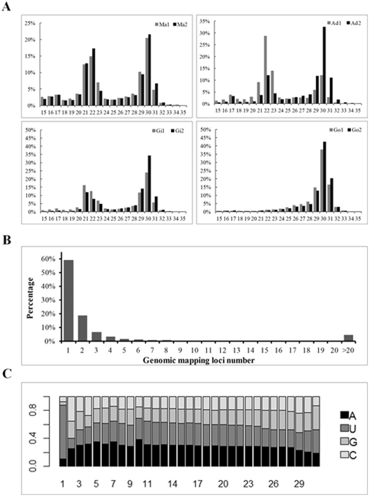
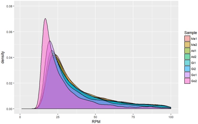
Piwi-interacting RNAs (piRNAs) belong to a recently discovered class of small non-coding RNAs whose best-understood function is repressing transposable element activity. Most piRNA studies have been conducted on model organisms and little is known about piRNA expression and function in mollusks. We performed high-throughput sequencing of small RNAs extracted from the mantle, adductor muscle, gill, and ovary tissues of the pearl oyster, Pinctada fucata. RNA species with sequences of approximately 30 nt were widely expressed in all tissues. Uridine at the 5' terminal and protection from β-elimination at the 3' terminal suggested that these were putative piRNAs. A total of 18.0 million putative piRNAs were assigned to 2.8 million unique piRNAs, and 35,848 piRNA clusters were identified. Mapping to the reference genome showed that 25% of the unique piRNAs mapped to multiple tandem loci on the scaffold. Expression patterns of the piRNA clusters were similar within the somatic tissues, but differed significantly between the somatic and gonadal tissues. These findings suggest that in pearl oysters piRNAs have important and novel functions beyond those in the germ line.


Conflict of interest statement

The authors declare no competing interests.

Figures

Figure 1
Figure 1
Putative piRNA sequence processing. (A). The size profile of small RNAs from the somatic and gonadal tissues of pearl oysters. The peak at 21–23 nt probably represents the microRNA and endogenous siRNA population. The peak at 26–31 nt probably represents putative piRNAs. Ma: mantle tissue; Ad: Adductor muscle; Gi: Gill tissue; Go: Gonad tissue. (B) Distribution of the number of loci at which each individual unique piRNA was mapped. A total of 0.82 million unique piRNAs with number of reads >1 were examined. (C) Base composition at each site of the unique piRNAs from the 5′ terminal.
Figure 2
Figure 2
Expression density of putative piRNAs from the somatic and gonadal tissues of pearl oysters. (A) Mantle libraries from two pearl oysters. (B) Adductor muscle libraries from two pearl oysters. (C) Gill libraries from two pearl oysters. (D). Gonad libraries from two pearl oysters. Ma: Mantle tissues; Ad: adductor muscle; Gi: Gill tissue; Go: Gonad tissue.
Figure 3
Figure 3
Results of mapping putative piRNAs to the reference genome. (A) Bidirectional mapping. (B,C) Unidirectional positive or negative strand mapping. The logs of piRNA mapping density (dark blue) and actual mapping for positive strand (light red) and negative strand (light blue) of the reference genome. The mapping density was plotted using the Integrative Genomics Viewer tracks tool.
Figure 4
Figure 4
Example of putative piRNA mapping according to multiple tandem loci on the scaffold. The dotted lines illustrate homologous sequence, which represent repeats on the reference genome.
Figure 5
Figure 5
β-elimination reaction of miRNA and putative piRNA oligonucleotides. (A) In the absence of 2′-O-methylation at the 3′ terminal, treated miR-279a (right) showed increased migration relative to untreated miR-279a (left) in a 15% polyacrylamide gel. (B) piRNA0001 showed the same migration before and after treatment, because methylation at the 3′ terminal protected it from periodate oxidation. F1 and F2: Female individuals total RNA; M1 and M2: male individuals total RNA. Gel figures are cropped and toned. The original gel images are available in Supplementary Information.
Figure 6
Figure 6
Detection of putative piRNAs in pearl oyster somatic tissues. (A) Northern blotting analysis of piRNA0001 expression in gill (Gi), adductor muscle (Ad), mantle (Ma), abdominal foot (Af), and intestine (In) tissues. (B) In situ hybridization analysis of piRNA0001 in mantle tissue. Bar = 50 μm. Gel figure is cropped and toned. The original gel image is available in Supplementary Information.

References

    1. Suzuki M, et al. An acidic matrix protein, Pif, is a key macromolecule for nacre formation. Science. 2009;325:1388–1390. doi: 10.1126/science.1173793. - DOI - PubMed
    1. Fang D, et al. Identification of genes directly involved in shell formation and their functions in pearl oyster. Pinctada fucata. PLoS One. 2011;6:e21860. doi: 10.1371/journal.pone.0021860. - DOI - PMC - PubMed
    1. Kinoshita S, et al. Deep sequencing of ESTs from nacreous and prismatic layer producing tissues and a screen for novel shell formation-related genes in the pearl oyster. PLoS One. 2011;6:e21238. doi: 10.1371/journal.pone.0021238. - DOI - PMC - PubMed
    1. Takeuchi T, et al. Draft genome of the pearl oyster Pinctada fucata: a platform for understanding bivalve biology. DNA Res. 2012;19:117–130. doi: 10.1093/dnares/dss005. - DOI - PMC - PubMed
    1. Mcginty EL, Evans BS, Taylor JUU, Jerry DR. Xenografts and pearl production in two pearl oyster species, P. maxima, and P. margaritifera: Effect on pearl quality and a key to understanding genetic contribution. Aquaculture. 2010;302:175–181. doi: 10.1016/j.aquaculture.2010.02.023. - DOI

Publication types

Substances