. 2019 Jan 8:9:3014.
doi: 10.3389/fmicb.2018.03014. eCollection 2018.

Structural Analysis of the Hanks-Type Protein Kinase YabT From Bacillus subtilis Provides New Insights in its DNA-Dependent Activation

Affiliations

Structural Analysis of the Hanks-Type Protein Kinase YabT From Bacillus subtilis Provides New Insights in its DNA-Dependent Activation

Lei Shi et al. Front Microbiol. .

Abstract

YabT is a serine/threonine kinase of the Hanks family from Bacillus subtilis, which lacks the canonical extracellular signal receptor domain but is anchored to the membrane through a C-terminal transmembrane helix. A previous study demonstrated that a basic juxtamembrane region corresponds to a DNA-binding motif essential for the activation of YabT trans-autophosphorylation. YabT is expressed during spore development and localizes to the asymmetric septum where it specifically phosphorylates essential proteins involved in genome maintenance, such as RecA, SsbA, and YabA. YabT has also been shown to phosphorylate proteins involved in protein synthesis, such as AbrB and Ef-Tu, suggesting a possible regulatory role in the progressive metabolic quiescence of the forespore. Finally, cross phosphorylations with other protein kinases implicate YabT in the regulation of numerous other cellular processes. Using an artificial protein scaffold as crystallization helper, we determined the first crystal structure of this DNA-dependent bacterial protein kinase. This allowed us to trap the active conformation of the kinase domain of YabT. Using NMR, we showed that the basic juxtamembrane region of YabT is disordered in the absence of DNA in solution, just like it is in the crystal, and that it is stabilized upon DNA binding. In comparison with its closest structural homolog, the mycobacterial kinase PknB allowed us to discuss the dimerization mode of YabT. Together with phosphorylation assays and DNA-binding experiments, this structural analysis helped us to gain new insights into the regulatory activation mechanism of YabT.

Keywords: autophosphorylation; crystallization chaperone; dimerization; regulatory mechanism; spore development.


Figures

Figure 1
Figure 1
Oligomerization state of the short and long forms of the bE8-YabT(∆TM) fusion protein. SEC-MALS analysis of the short form injected at 3 mg/ml (A) and of the long form injected at 4 mg/ml (B). The short form was eluted as a single peak corresponding to a dimer of about 115 kDa. The long form was eluted as two main peaks, which were analyzed as an equilibrium between a monomer of about 60 kDa and a dimer of about 120 kDa. An additional small peak suggested the formation of a tetramer. The two main peaks were analyzed separately but are displayed on the same graph. The elution profiles are represented according to retention volume (in ml) with the refractive index (in mV) indicated on the left axis (black curve) and the logarithm of molecular weight on the right axis (red curve). (C) Models of the intra- and inter-molecular interactions of the bE8 and YabT(∆TM) domains of the fusion proteins. The linker between bE8 and the bilobal kinase domain is shown in red for the short form and in green for the long form. The specific contacts between bE8 and YabT are shown as dashed lines. A subunit is colored in dark blue and the other one in pale blue.
Figure 2
Figure 2
DNA-binding activity of the short and long forms of the bE8-YabT(∆TM) fusion protein. 0.075 μM dsDNA (210 bp) (A) and 0.15 μM ssDNA (90 nt) (B) were incubated at 310 K for 30 min with 0 μM (lane 1, 5 and 9), 0.4 μM (lane 2, 6 and 10), 0.8 μM (lane 3, 7 and 11), and 1.2 μM (lane 4, 8 and 12) of protein, respectively. The samples were then submitted to electrophoretic mobility shift assays (EMSA). The activity of YabT(∆TM) is used as positive control.
Figure 3
Figure 3
Autophosphorylation activity of the short and long forms of the bE8-YabT(∆TM) fusion protein. About 1.6 μM protein samples were incubated with 0 nM (lane 1, 3 and 6), 33 nM (lanes 2, 4, and 7), and 67 nM (lanes 5 and 8) dsDNA. Autophosphorylation reactions were started by adding 10 mM ATP containing 20 mCi mmol-1 [γ-32P]-ATP. The samples were analyzed by SDS-PAGE, and the radioactive phosphorylated proteins were revealed by autoradiography. The activity of the short and long forms of the bE8-YabT(∆TM) fusion protein is compared to the activity of a YabT(∆TM) sample used as positive control. Phosphorylated YabT and bE8-YabT samples are indicated by arrows. Upper band(s) are observed when YabT is active. They could correspond either to minor multimeric states of the phosphorylated protein or to a minor E. coli contaminant recognized as substrate by the poorly specific YabT.
Figure 4
Figure 4
Structure of the bE8-YabT(∆TM) fusion proteins. (A) Overall structure of the bE8-YabT(∆TM) monomeric form. The YabT(∆TM) is shown in cartoon format and the bE8 αREP binder as ribbon. Both domains are colored in a rainbow scheme from blue (N-terminus) to red (C-terminus). The secondary structure elements of YabT(∆TM) are labeled. The linker region is not visible, but both domains display the characteristic αREP interaction mode. The distance of 60 Å separating the C-terminus of bE8 from the N-terminus of YabT is highlighted by a dashed line. It is compatible with a 32 residues long linker. (B) Close view of the bE8/YabT(∆TM) interaction. The bE8 surface is shown in transparency. YabT residues directly implicated in polar interactions with bE8 are labeled and shown in sticks and dots. (C) Alternative dimeric form of the bE8-YabT(∆TM) fusion protein observed in crystal packing. A subunit is shown in the same scheme as in panel A. The second subunit is shown in gray. In each subunit, the C-terminus of the bE8 domain is only 20 Å apart from the N-terminus of the YabT(∆TM) domain but specifically interact with the YabT(∆TM) domain of the second subunit. (D) Characteristics of the active closed conformation. The electrostatic interactions made by Lys55 from strand β3 and residues Lys173 and Arg176 from the activation loop are highlighted by dashed lines. The hydrophobic interactions between Val168 from the activation loop (yellow) and helix αC (cyan) are highlighted by dots. A neighboring bE8 molecule from the crystal packing is shown as gray ribbon with residues Arg21*, Asp46*, and Glu47* in sticks.
Figure 5
Figure 5
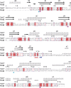
Sequence alignment between YabT and its closest structural homolog PknB from Mycobacterium tuberculosis. The secondary structure elements are shown at the top, using the PDB entry 2FUM for PknB. YabT residues discussed in the text are highlighted by logos at the bottom of the alignment: F146G147D148 and D167V168G169 motifs as red triangles, regulatory autophosphorylation sites T171 and T172 as blue spheres, and PtkA phosphorylation sites Y28, Y92, and Y254 as magenta squares. Figure prepared using ESPript (http://espript.ibcp.fr) (Robert and Gouet, 2014).
Figure 6
Figure 6
NMR analysis of YabT in the presence and absence of DNA. Superimposition of the Sofast 1H-15N HMQC NMR spectra of isotopically labeled 6His-YabT(∆TM) alone (in gray) (A), after addition of ATP and DNA (in magenta), and (B), after addition of ATP alone (in green). Each peak represents a bonded N-H pair, with its two coordinates corresponding to the chemical shifts of each of the H and N atoms. Only the region of the spectra corresponding to disordered residues is shown. The peaks corresponding to glycine residues are circled by dashed lines.
Figure 7
Figure 7
Comparison of the bE8-YabT(∆TM) structure with the two types of PknB dimerization modes. YabT(∆TM) is shown as cartoon colored in rainbow scheme. bE8 is shown as ribbon colored in grey. The PknB subunits are shown as cartoon colored in beige. The surface of the PknB subunit, which is not superimposed on YabT(∆TM), is shown in transparency. (A) Superimposition with the back-to-back dimer of PknB (PDB ID 2FUM). (B) Superimposition with the face-to-face PknB dimer (PDB ID 3F69).
Figure 8
Figure 8
Model of a face-to-face YabT(∆TM) dimer. The model was built by superimposition of the structure of YabT(∆TM) on both subunits of the face-to-face dimer of PknB (PDB ID 3F69). The electrostatic surface of the dimer is shown in transparency with negatively and positively charged region colored in red and blue, respectively. Each polypeptide chain is shown as a cartoon trace colored in a rainbow scheme from blue to red. Tyrosine residues phosphorylated by the BY-kinase PtkA are highlighted by dots and labeled.

Similar articles

Cited by

References

    1. Adams P. D., Afonine P. V., Bunkoczi G., Chen V. B., Davis I. W., Echols N., et al. . (2010). PHENIX: a comprehensive Python-based system for macromolecular structure solution. Acta Crystallogr. D. Biol. Crystallogr. 66, 213–221. 10.1107/S0907444909052925, PMID: - DOI - PMC - PubMed
    1. Alber T. (2009). Signaling mechanisms of the Mycobacterium tuberculosis receptor Ser/Thr protein kinases. Curr. Opin. Struct. Biol. 19, 650–657. 10.1016/j.sbi.2009.10.017, PMID: - DOI - PMC - PubMed
    1. Barthe P., Mukamolova G. V., Roumestand C., Cohen-Gonsaud M. (2010). The structure of PknB extracellular PASTA domain from Mycobacterium tuberculosis suggests a ligand-dependent kinase activation. Structure 18, 606–615. 10.1016/j.str.2010.02.013, PMID: - DOI - PubMed
    1. Bidnenko V., Shi L., Kobir A., Ventroux M., Pigeonneau N., Henry C., et al. . (2013). Bacillus subtilis serine/threonine protein kinase YabT is involved in spore development via phosphorylation of a bacterial recombinase. Mol. Microbiol. 88, 921–935. 10.1111/mmi.12233, PMID: - DOI - PMC - PubMed
    1. Bocharov E. V., Sharonov G. V., Bocharova O. V., Pavlov K. V. (2017). Conformational transitions and interactions underlying the function of membrane embedded receptor protein kinases. Biochim. Biophys. Acta 1859, 1417–1429. 10.1016/j.bbamem.2017.01.025 - DOI - PubMed

LinkOut - more resources