. 2019 Jan 8:9:3184.
doi: 10.3389/fmicb.2018.03184. eCollection 2018.

Discovery and Characterization of Mycobacterium basiliense sp. nov., a Nontuberculous Mycobacterium Isolated From Human Lungs

Affiliations

Discovery and Characterization of Mycobacterium basiliense sp. nov., a Nontuberculous Mycobacterium Isolated From Human Lungs

Helena M B Seth-Smith et al. Front Microbiol. .

Abstract

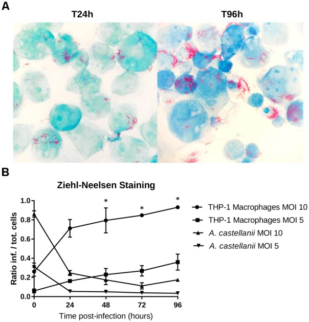
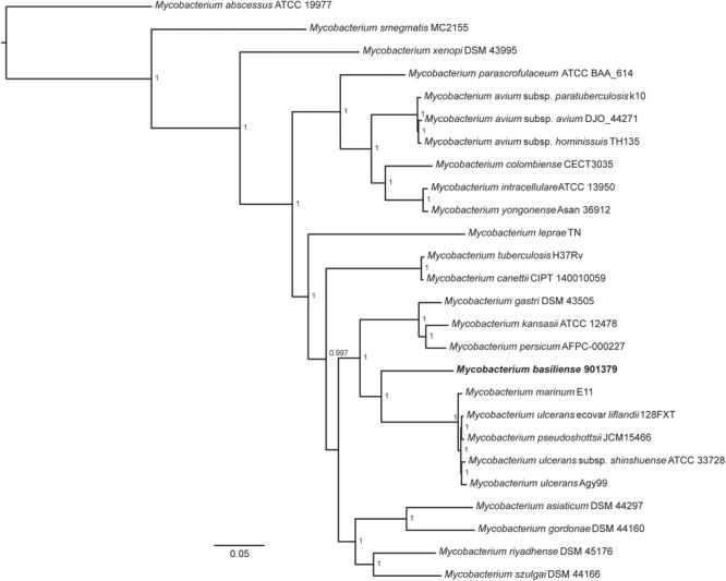
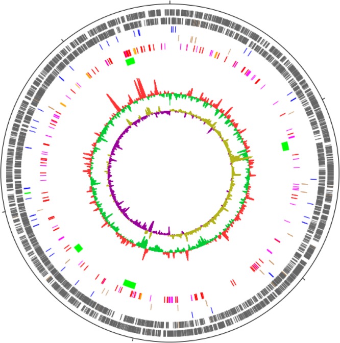
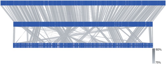
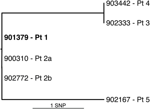
Bacteria belonging to the genus Mycobacterium are predominantly responsible for pulmonary diseases; most notably Mycobacterium tuberculosis causes granulomatous pulmonary infections. Here we describe a novel slow growing mycobacterial species isolated from respiratory samples from five patients, four with underlying pulmonary disease. The isolates were characterized by biochemical and molecular techniques, including whole genome sequencing. Biochemical characteristics generally match those of M. marinum and M. ulcerans; however, the most striking difference of the new species is its ability to grow at 37°C. The new species was found to grow in human macrophages, but not amoebae, suggesting a pathogenic rather than an environmental lifestyle. Phylogenetic analysis reveals a deep-rooting relationship to M. marinum and M. ulcerans. A complete genome sequence was obtained through combining short and long-read sequencing, providing a genome of 5.6 Mb. The genome appears to be highly intact, syntenic with that of M. marinum, with very few insertion sequences. A vast array of virulence factors includes 283 PE/PPE surface-associated proteins, making up 10% of the coding capacity, and 22 non-ribosomal peptide synthase clusters. A comparison of six clinical isolates from the five patients shows that they differ by up to two single nucleotide polymorphisms, suggesting a common source of infection. Our findings are in accordance with the recognition of a new taxonomic entity. We propose the name M. basiliense, as all isolates were found in patients from the Basel area of Switzerland.

Keywords: Mycobacterium basiliense; nontuberculous mycobacteria; novel species; pathogen; virulence.


Figures

FIGURE 1
FIGURE 1
Representative high-performance liquid chromatography (HPLC) patterns showing cell-wall mycolic acid content of Mycobacterium basiliense in comparison with M. marinum and M. tuberculosis. Numbers indicate retention times (minutes). ITS, internal standard.
FIGURE 2
FIGURE 2
Growth in THP-1 human macrophages and in Acanthamoeba castellanii ATCC 30010. (A) DNA copies/ml over the growth time (means ± 2 standard deviations). (B) CFU/ml over the growth time (means ± 2 standard deviations).
FIGURE 3
FIGURE 3
THP-1 macrophage infection shown using Ziehl-Neelsen staining. (A) THP-1 cells strained with ZN 24 and 96 h post-infection with an MOI of 10. Mycobacteria were observed in the extracellular medium, probably explained by recent host cell lysis events; extracellular growth is unlikely to have occurred because infected cells were treated with gentamycin to remove bacteria that were not internalized. (B) Ratio of the infected cells to the total number of cells counted (means ± 2 standard deviations). The asterisks () indicate the timepoints at which each condition was significantly different from another by doing pair-wise chi-squared tests between pairs of triplicates with five degrees of freedom (p-value < 0.05).
FIGURE 4
FIGURE 4
Core genome phylogeny of slow growing mycobacterial species. M. basiliense can be seen to be most closely related to M. ulcerans, M. marinum, M. ulcerans ecovar liflandii and M. pseudoshottsii, as well as to the M. kansasii complex. Shared core genes (n = 768) were obtained through clustering CDSs into orthologous groups using OrthoFinder v2.2.1 (Emms and Kelly, 2015). Single-copy orthologous genes were aligned using Mafft v7.310 (Katoh and Standley, 2013), resulting in a concatenated alignment of 283,167 amino acids. A maximum likelihood tree was built using FastTree v2.1.10 Double precision using parameters (–gamma –spr 4 –mlacc 2) (Price et al., 2010). The tree was rooted on M. abscessus ATCC 19977 using Archaeopteryx 0.9921 (Zmasek, 2015) (https://sites.google.com/site/cmzmasek/home/software/archaeopteryx). The scale bar represents the number of amino acids substitutions per site alongside the branches. Nodes supports are based on the Shimodaira-Hasegawa (SH) test. NB, BLAST (using BLASTn) of the M. shottsii-specific F5 fragment (accession number HM149249) against the M. basiliense assembly returned no hit.
FIGURE 5
FIGURE 5
Circular map of the draft genome of M. basiliense strain 901379. The 5.6 Mb chromosome is represented as a circle, with ticks every 1 Mb. The circles represent, from outside to inside: CDSs encoded on the forward strand; CDSs encoded on the reverse strand; tRNAs (blue) and rRNAs (green); pseudogenes (brown); CDSs encoding PE (red), PPE (pink) and Mce (orange) proteins; regions not present in M. marinum strain M (green); G+C content; G+C skew. Figure was produced in DNAPlotter (Carver et al., 2009).
FIGURE 6
FIGURE 6
Comparison of the genomes of M. basiliense 901379 (middle) with M. marinum M (CP000854, top) and M. ulcerans Agy99 (CP000325, bottom). Genomes are depicted linearly with predicted CDSs as blue vertical lines. Blastn hits between the genomes (identity shown in scale) show that the genome of M. basiliense is syntenic with that of M. marinum, whereas the genome of M. ulcerans contains many rearrangements. Figure was drawn using Easyfig (Sullivan et al., 2011).
FIGURE 7
FIGURE 7
Genome phylogeny of six M. basiliense isolates. SNP phylogeny of the full genome based on high quality, manually checked SNPs. Root is assigned to the type strain (earliest isolate, bold), which was also the mapping reference. Additional isolates, identified by isolate number and patient, vary by a maximum of two SNPs from the root.

References

    1. Auch A., Von Jan M., Klenk H.-P., Göker M. (2010). Digital DNA-DNA hybridization for microbial species delineation by means of genome-to-genome sequence comparison. Stand. Genomic Sci. 2 117–134. 10.4056/sigs.531120 - DOI - PMC - PubMed
    1. Boddinghaus B., Rogall T., Flohr T., Blocker H., Bottger E. C. (1990). Detection and identification of mycobacteria by amplification of rRNA. J. Clin. Microbiol. 28 1751–1759. - PMC - PubMed
    1. Brown-Elliott B. A., Wallace R. J., Jr. (2002). Clinical and taxonomic status of pathogenic nonpigmented or late-pigmenting rapidly growing mycobacteria. Clin. Microbiol. Rev. 15 716–746. 10.1128/CMR.15.4.716-746.2002 - DOI - PMC - PubMed
    1. Carver T. J., Rutherford K. M., Berriman M., Rajandream M. A., Barrell B. G., Parkhill J. (2005). ACT: the artemis comparison tool. Bioinformatics 21 3422–3423. 10.1093/bioinformatics/bti553 - DOI - PubMed
    1. Carver T., Thomson N., Bleasby A., Berriman M., Parkhill J. (2009). DNAPlotter: circular and linear interactive genome visualization. Bioinformatics 25 119–120. 10.1093/bioinformatics/btn578 - DOI - PMC - PubMed