. 2018 Dec;13(4):115-121.
doi: 10.1016/j.joto.2018.08.002. Epub 2018 Aug 30.

Cisplatin-induced vestibular hair cell lesion-less damage at high doses

Affiliations

Cisplatin-induced vestibular hair cell lesion-less damage at high doses

Dalian Ding et al. J Otol. 2018 Dec.

Erratum in

Abstract

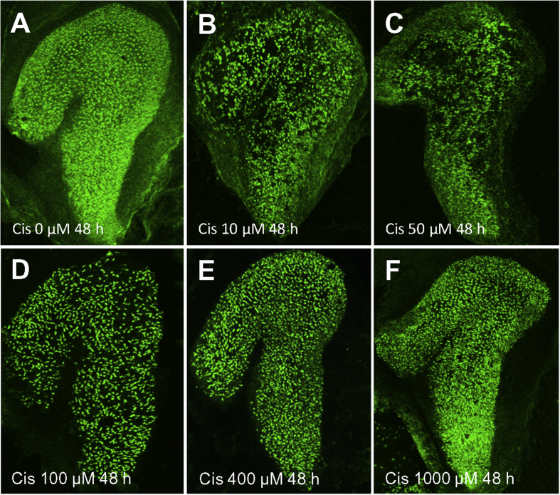
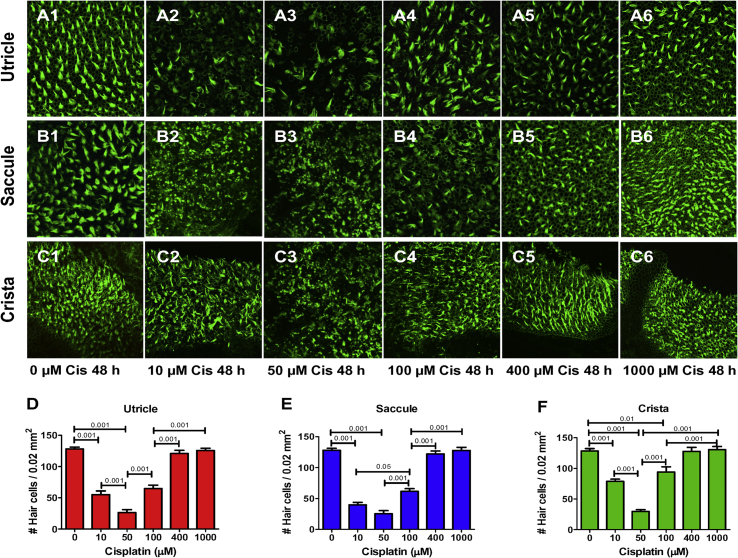
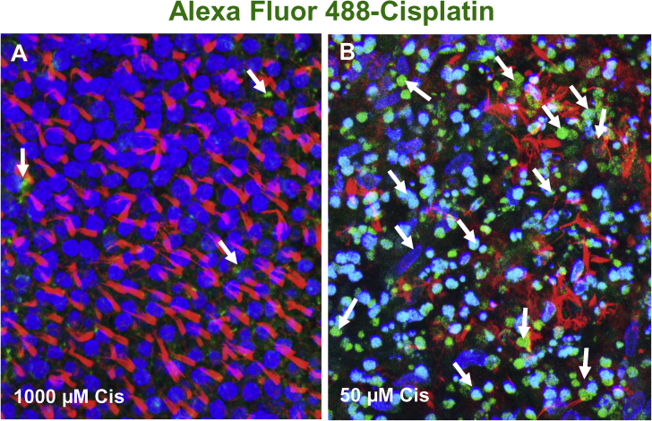
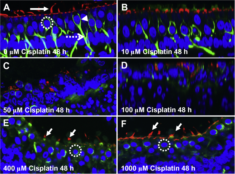
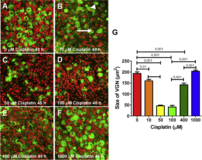
Cisplatin, a widely used anticancer drug, damages hair cells in cochlear organotypic cultures at low doses, but paradoxically causes little damage at high doses resulting in a U-shaped dose-response function. To determine if the cisplatin dose-response function for vestibular hair cells follows a similar pattern, we treated vestibular organotypic cultures with doses of cisplatin ranging from 10 to 1000 μM. Vestibular hair cell lesions progressively increased as the dose of cisplatin increased with maximum damage occurring around 50-100 μM, but the lesions progressively decreased at higher doses resulting in little hair cell loss at 1000 μM. The U-shaped dose-response function for cisplatin-treated vestibular hair cells in culture appears to be regulated by copper transporters, Ctr1, ATP7A and ATP7B, that dose-dependently regulate the uptake, sequestration and extrusion of cisplatin.

Keywords: Cisplatin; Copper transporters; Ototoxicity; Vestibular organotypic cultures.


Figures

Fig. 1
Fig. 1
Photomicrographs showing the whole macula of saccule after 48 h treatment (A) without (control) and with cisplatin (Cis) at concentrations of 10–1000 μM (B–F). Specimens stained with Alexa 488-conjugated phalloidin which preferentially labels the stereocilia bundles and cuticular plate of vestibular hair cells. (A) Note high density of hair cell stereocilia in specimen cultured without cisplatin for 48 h. (B–D) Many hair cells were missing after treatment with 10, 50, or 100 μM cisplatin for 48 h. (E–F) Hair cell density in cultures treated with 400 or 1000 μM cisplatin were greater than with 10 or 50 μM cisplatin.
Fig. 2
Fig. 2
Representative photomicrographs of vestibular explants from utricle, saccule and crista cultured for 48 h without cisplatin (Cis) or with 10, 50, 100, 400 or 1000 μM cisplatin. Specimens stained with Alexa 488-conjugated phalloidin which labels the hair cell stereocilia bundles. Macula of utricle (A1), macula of saccule (B1) and ampulla of crista (C1) cultured with 0 μM cisplatin for 48 h; note high density of stereocilia bundles in control cultures. Density of hair cell stereocilia reduced at cisplatin concentrations of 10–50 μM in utricle (A2-3), saccule (B2-3) and crista (C2-3), but hair cell density gradually increased as the concentration increased from 100 to 1000 μM (A4,5,6; B4,5,6; C4,5,6). D–F: Histograms showing mean (n = 6, +/−) number of hair cells per 0.22 μm2 in utricle, saccule and ampula. Horizontal lines with p values above indicate significant differences between conditions (p < 0.05, 0.01 or 0.001, one-way ANOVA, Bonferroni post-hoc comparisons).
Fig. 3
Fig. 3
Photomicrographs showing accumulation of Alexa Fluor 488-conjugated cisplatin (green near arrows) in hair cells in the macula of utricle. Nuclei labeled with ToPro-3 (blue); hair cell stereocilia labeled with Alex Fluor 555-conjugated phalloidin (red). (A) In cultures treated with 1000 μM cisplatin, stereocilia bundles (red) were arranged in tufts and ToPro-3 nuclei were large and round, morphological features of healthy hair cells. Only a few puncta of Alex 488-cisplatin (arrow) present in hair cells. (B) In cultures treated with 50 μM cisplatin, extensive Alexa 488-cisplatin (green/turquoise) labeling was evident in vestibular hair cells. The stereocilia bundles (red) were either missing or in disarray and many hair cell nuclei were condensed (blue/turquoise) indicative of cells undergoing apoptosis.
Fig. 4
Fig. 4
Representative Z-plane photomicrographs showing uptake of AM1-43 (red) into vestibular hair cells (HC) in macula of utricle. Nuclei of cells labeled with ToPro-3 (blue) to label nuclei and Alex Fluor 488-conjugated phalloidin (green) to identify hair cell stereocilia and cuticular plate. (A) In controls (0 μM cisplatin), robust AM1-43 labeling was present in hair cell cytoplasm, but was absent from support cells (SC) beneath the hair cells. (B) After 48 h treatment with 10 μM cisplatin, AM1-43 was reduced or absent from hair cells with damaged or missing stereocilia (arrowhead). After 48 h treatment with 50 μM cisplatin, many hair cells were damaged (dotted circles around shrunken nuclei) and only small, scattered puncta of AM1-43 were present in damaged areas of the epithelium. (D) After 48 h treatment with 1000 μM cisplatin, most hair cells were intact (large nuclei, phalloidin label on apical surface of hair cells); however, AM1-43 was absent from hair cells. (E) Cultures treated for 18 h with 50 μM cisplatin; then culture medium was replaced with standard culture medium and cultured an additional 18 h after which AM1-43 was added to medium. Note damage to stereocilia and shrunken nuclei (dotted circle) due to low-dose cisplatin. (F) Same conditions as E except cisplatin dose was 1000 μM cisplatin. Note robust AM1-43 labeling in cytoplasm and phallodin labeling of cuticular plate of hair cells, which appear normal.
Fig. 5
Fig. 5
Photomicrographs of vestibular hair cells and nerve endings. (A) Control vestibular explant cultured for 48 h. Stereocilia and cuticular plate labeled with Alexa Fluor 555-phalloidin (red, arrow), nuclei labeled with ToPro-3 (blue) and nerve fibers labeled with beta-tubulin (green). Note chalice shaped afferent ending (arrowhead) around hair cells, afferent nerve fiber (dashed arrow) and large round nucleus (dotted circle) of hair cells. (B): After 48 h treatment with 10 μM cisplatin, no major changes observed in ToPro-3 labeled nuclei, phalloidin labeled hair cells or tubulin-labeled nerve fibers. (C–D) After 48 h treatment with 50 and 100 μM cisplatin treatment, most vestibular hair and afferent nerve fibers missing. (E) In vestibular explant treated with 400 μM cisplatin for 48 h, most hair cell nuclei present, but nuclei generally smaller than controls. Nerve fibers missing and chalice afferent terminals largely absent. (F) Note phalloidin labeling of apical pole of hair cell and stereocilia and round nuclei of sensory hair cell (dotted circle) after 48 treatment with 1000 μM cisplatin. Nerve fibers missing and chalice shaped afferent terminal shrunken or largely absent.
Fig. 6
Fig. 6
Vestibular ganglion neurons (VGN) cultured for 48 h without (A) (0 μm) or with (B–F) 10, 50, 100, 400 and 1000 μM cisplatin respectively. Samples labeled with β−tubulin III (green) to label neurons and ToPro-3 (red) to label nuclei. (A) Note large round soma and round nucleolus in the absence of cisplatin. (B) Treatment with 10 μM cisplatin resulted shrinkage of nucleolus (arrow) and soma (arrowhead) of some neurons. (C–D) Treatment with 50 and 100 μM cisplatin resulted in soma loss, and shrinkage, condensation and fragmentation of the nucleolus. (E–F) Note increases in the number of neurons and size of the soma and nucleolus as the cisplatin dose increased from 400 to 1000 μM. (G) Mean size of vestibular ganglion soma initially decreased as the cisplatin dose increased from zero to 100 μM cisplatin, but soma size incresed as the cisplatin dose increased from 400 to 1000 μM. Horizontal bars show conditions that were significantly different from one another (Bonferroni p values shown above bar).

References

    1. Alam S.A., Ikeda K., Oshima T., Suzuki M., Kawase T., Kikuchi T., Takasaka T. Cisplatin-induced apoptotic cell death in Mongolian gerbil cochlea. Hear. Res. 2000;141:28–38. - PubMed
    1. Cepero V., Garcia-Serrelde B., Moneo V., Blanco F., Gonzalez-Vadillo A.M., Alvarez-Valdes A., Navarro-Ranninger C., Carnero A. Trans-platinum(II) complexes with cyclohexylamine as expectator ligand induce necrosis in tumour cells by inhibiting DNA synthesis and RNA transcription. Clin. Transl. Oncol. 2007;9:521–530. - PubMed
    1. Chen Y., Heeg M.J., Braunschweiger P.G., Xie W., Wang P.G. A carbohydratee-linked cisplatin analogue having antitumor activity. Angew. Chem. Int. Ed. 1999;38:1768–1769. - PubMed
    1. Ding D., Allman B.L., Salvi R. Review: ototoxic characteristics of platinum antitumor drugs. Anat. Rec. 2012;295:1851–1867. - PubMed
    1. Ding D., Allman B.L., Yin S., Sun H., Salvi R. Cisplatin ototoxicity. In: Dupont J., editor. Hearing Loss: Classification, Causes and Treatment. Nova Science Publishers, Inc.; Hauggauge, NY: 2011. pp. 39–63.