

doi: 10.1177/1941874418758902.
Epub 2018 Feb 20.
An Unusual Etiology of Acute Spontaneous Intracerebral Hemorrhage
Affiliations
- PMID: 30671164
- PMCID: PMC6327245
- DOI: 10.1177/1941874418758902
Item in Clipboard
An Unusual Etiology of Acute Spontaneous Intracerebral Hemorrhage
Neurohospitalist.
2019 Jan.
No abstract available
Keywords: central nervous system infections; cerebrovascular disorders; epilepsy; intracranial hemorrhages; neuropathology; seizures; status epilepticus; techniques.
Conflict of interest statement
Declaration of Conflicting Interests: The authors declared no potential conflicts of interest with respect to the research, authorship, and/or publication of this article.
Figures
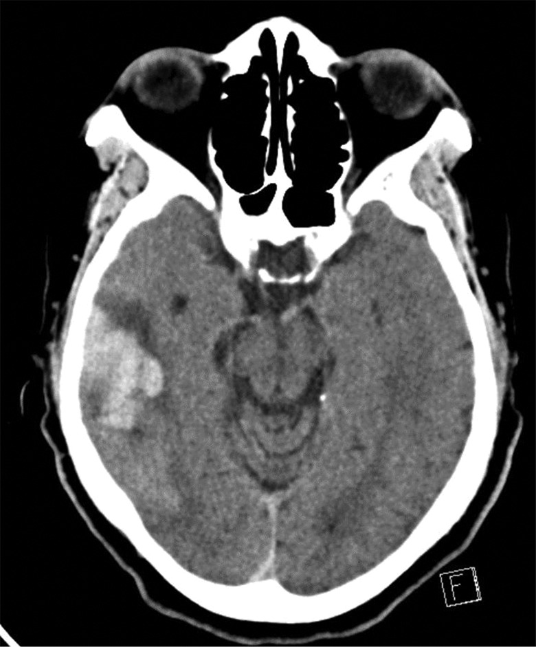
Initial noncontrast head computed tomography demonstrating a 2.5 × 3.2 cm right lateral
temporal lobe intracerebral hemorrhage.
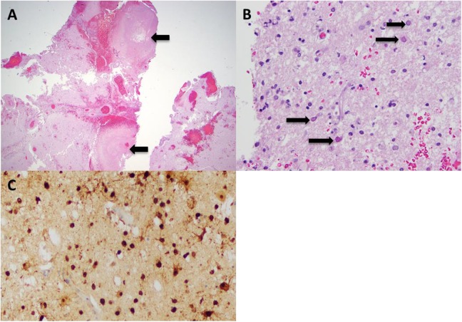
Histopathological findings from right temporal lobe tissue. A, Low-power (×40)
hematoxylin–eosin stain of right temporal lobe tissue showing hypercellular parenchyma
with lymphoplasmacytic and neutrophilic infiltrate as well as hemorrhage and necrosis
(arrows). B, High-power (×500) hematoxylin–eosin stain showing astrocytes with
cytopathic effect and intranuclear inclusions (arrows). C, Immunohistochemical staining
(×200) showing strong diffuse positivity for herpes simplex virus 1 (HSV-1).
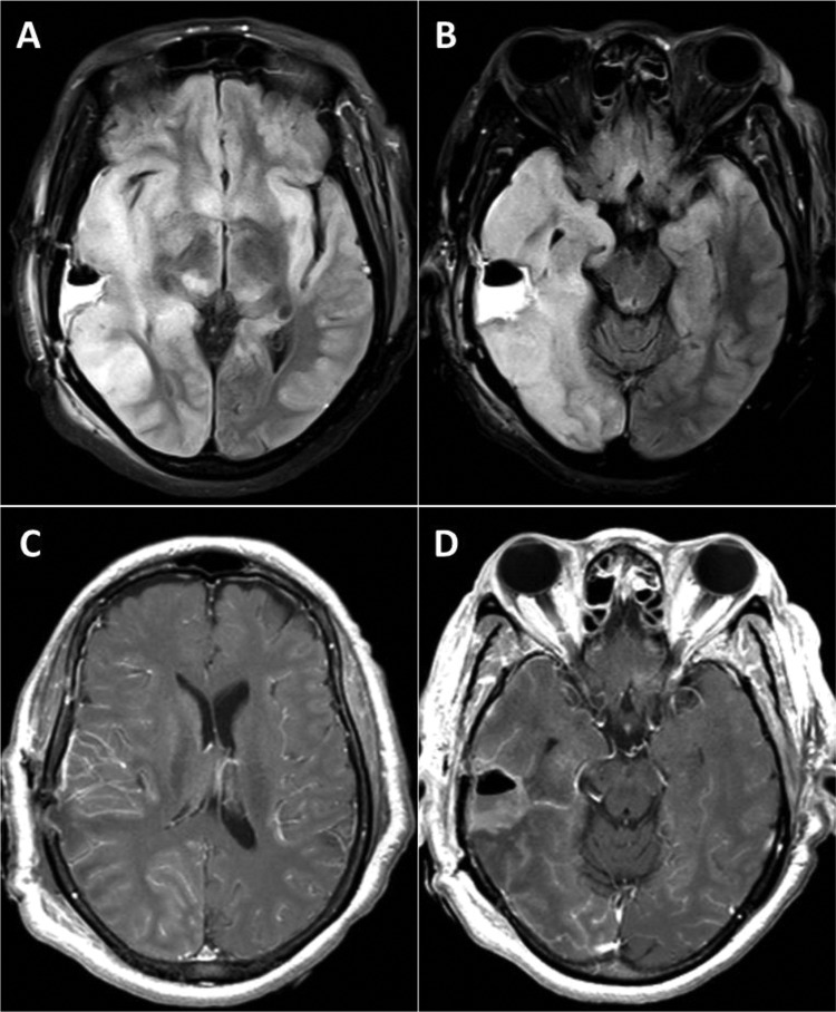
Postoperative brain magnetic resonance imaging (MRI) findings. A and B, Postoperative
axial MRI of the brain showing diffuse T2 fluid-attenuated inversion recovery signal
hyperintensities in the bilateral hemispheres involving cortical and subcortical
structures. C and D, Postoperative axial MRI of the brain showing diffuse gyral
gadolinium enhancement throughout the right parietal and temporal lobes.
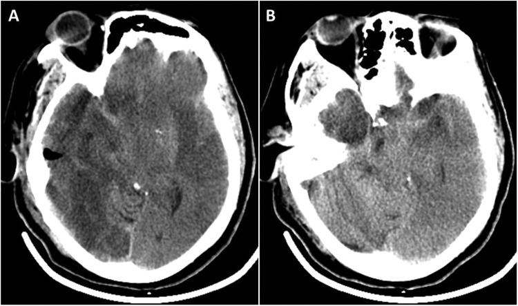
A and B, Final noncontrast head computed tomography demonstrating diffuse hypodensities
throughout right and left hemisphere with complete effacement of the basal cisterns and
uncal herniation.
Similar articles
-
Intracranial Calcification Masquerading as Hemorrhage in a Patient with Multiple Myeloma Presenting with Facial Neuropathy.Cureus. 2018 Jun 19;10(6):e2841. doi: 10.7759/cureus.2841. Cureus. 2018. PMID: 30131933 Free PMC article.
-
Seizures and CNS hemorrhage: spontaneous intracerebral and aneurysmal subarachnoid hemorrhage.Neurologist. 2010 May;16(3):165-75. doi: 10.1097/NRL.0b013e3181c7cd0b. Neurologist. 2010. PMID: 20445426 Review.
-
Suppressing cAMP response element-binding protein transcription shortens the duration of status epilepticus and decreases the number of spontaneous seizures in the pilocarpine model of epilepsy.Epilepsia. 2015 Dec;56(12):1870-8. doi: 10.1111/epi.13211. Epub 2015 Sep 30. Epilepsia. 2015. PMID: 26419901 Free PMC article.
-
[Spontaneous intracerebral hemorrhage: the clinical neuroradiological view].Radiologe. 1999 Oct;39(10):828-37. doi: 10.1007/s001170050719. Radiologe. 1999. PMID: 10550381 Review. German.
-
Does a Spontaneous Intracerebral Hemorrhage Predispose to a Secondary, Distant Intracerebral Hemorrhage? A Case Report and Review of the Literature.Cureus. 2017 Dec 29;9(12):e1999. doi: 10.7759/cureus.1999. Cureus. 2017. PMID: 29507847 Free PMC article.
Cited by
-
Cerebrovascular manifestations of herpes simplex virus infection of the central nervous system: a systematic review.J Neuroinflammation. 2019 Jan 29;16(1):19. doi: 10.1186/s12974-019-1409-4. J Neuroinflammation. 2019. PMID: 30696448 Free PMC article.
-
Potential mechanisms of hemorrhagic stroke in elderly COVID-19 patients.Aging (Albany NY). 2020 Jun 11;12(11):10022-10034. doi: 10.18632/aging.103335. Epub 2020 Jun 11. Aging (Albany NY). 2020. PMID: 32527987 Free PMC article. Review.
-
Limited efficacy of adjunctive therapy in a case of severe herpes simplex virus encephalitis: A case report and literature review.IDCases. 2025 Jun 26;41:e02307. doi: 10.1016/j.idcr.2025.e02307. eCollection 2025. IDCases. 2025. PMID: 40688426 Free PMC article.
-
A Rare Presentation of Herpes Simplex Virus Encephalitis With Ischemic Stroke and Associated Acyclovir-Induced Acute Kidney Injury.Cureus. 2024 Oct 20;16(10):e71978. doi: 10.7759/cureus.71978. eCollection 2024 Oct. Cureus. 2024. PMID: 39569230 Free PMC article.
References
-
- Qureshi AI, Tuhrim S, Broderick JP, Batjer HH, Hondo H, Hanley DF. Spontaneous intracerebral hemorrhage. N Engl J Med. 2001;344(19):1450–1460. doi:10.1056/NEJM200105103441907. - PubMed
-
- Thomas B, Krishnamoorthy T, Purkayastha S, Gupta AK. Isolated left vein of Labbe thrombosis. Neurology. 2005;65(7):1135 doi:10.1212/01.wnl.0000181352.14678.18. - PubMed
-
- Kimura K, Iguchi Y, Inoue T, et al. Hyperglycemia independently increases the risk of early death in acute spontaneous intracerebral hemorrhage. J Neurol Sci. 2007;255(1-2):90–94. doi:10.1016/j.jns.2007.02.005. - PubMed
-
- Behrouz R, Hafeez S, Miller CM. Admission leukocytosis in intracerebral hemorrhage: associated factors and prognostic implications. Neurocritical Care. 2015;23(3):370–373. doi:10.1007/s12028-015-0128-7. - PubMed
LinkOut - more resources
Full Text Sources