. 2019 Jan 16:7:e6284.
doi: 10.7717/peerj.6284. eCollection 2019.

CRISPR/Cas9-mediated deletion of the Wiskott-Aldrich syndrome locus causes actin cytoskeleton disorganization in murine erythroleukemia cells

Affiliations

CRISPR/Cas9-mediated deletion of the Wiskott-Aldrich syndrome locus causes actin cytoskeleton disorganization in murine erythroleukemia cells

Vanessa Fernández-Calleja et al. PeerJ. .

Abstract

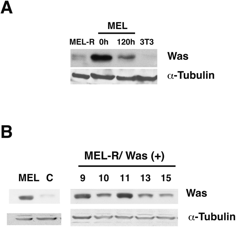
Wiskott-Aldrich syndrome (WAS) is a recessive X-linked inmmunodeficiency caused by loss-of-function mutations in the gene encoding the WAS protein (WASp). WASp plays an important role in the polymerization of the actin cytoskeleton in hematopoietic cells through activation of the Arp2/3 complex. In a previous study, we found that actin cytoskeleton proteins, including WASp, were silenced in murine erythroleukemia cells defective in differentiation. Here, we designed a CRISPR/Cas9 strategy to delete a 9.5-kb genomic region encompassing the Was gene in the X chromosome of murine erythroleukemia (MEL) cells. We show that Was-deficient MEL cells have a poor organization of the actin cytoskeleton that can be recovered by restoring Was expression. We found that whereas the total amount of actin protein was similar between wild-type and Was knockout MEL cells, the latter exhibited an altered ratio of monomeric G-actin to polymeric F-actin. We also demonstrate that Was overexpression can mediate the activation of Bruton's tyrosine kinase. Overall, these findings support the role of WASp as a key regulator of F-actin in erythroid cells.

Keywords: Actin cytoskeleton; Bruton tyrosine kinase; CRISPR/Cas9; Erythroleukemia cells; Wiskott-Aldrich.


Conflict of interest statement

Dora B. Krimer is an Academic Editor for PeerJ.

Figures

Figure 1
Figure 1. WASp is poorly expressed in MEL-R cells.
(A) Immunoblot analysis of whole-cell extracts from erythroleukemia-resistant cells (MEL-R), MEL cells undifferentiated (0 h) and differentiated with hexamethylene bisacetamide (HMBA) (120 h), and 3T3 fibroblasts. Equal amounts of protein (30 µg) were fractionated by SDS-polyacrylamide gel electrophoresis and analyzed by immunoblotting with an anti-Was antibody. α-tubulin was used as a loading control. (B) Immunoblot analysis of whole-cell lysates from stable transfectants overexpressing Was (MEL-R/Was (+) processed as in A). Numbers above the panel correspond to clones 8, 9, 10, 11, 13 and 15. MEL cells and MEL-R cells transfected with an empty vector (C) were treated and analyzed under similar conditions. α-tubulin was used as a loading control.
Figure 2
Figure 2. Overexpression of Was. induces organization and polymerization of actin cytoskeleton in MEL-R cells.
Immunofluorescence staining of MEL cells, MEL-R/Was (+) transfectants 9, 10 and 11, and MEL-R cells with a mouse monoclonal anti-b-actin antibody (red). Nuclei were visualized with DAPI (blue). Magnified views indicated by white boxed areas are shown below second-row panels. The scale bar represents 10 mm.
Figure 3
Figure 3. Overexpression of Was enhances the formation of F-actin in MEL-R transfectants.
(A) Total actin expression was evaluated in MEL-R/Was (+) clones 9, 10 and 11, and MEL-R cells by immunoblotting with an antibody against b-actin. α-tubulin was used as a loading control. (B) G-actin and F-actin from MEL-R and MEL-R/Was (+) transfectants 9, 10 and 11, separated after ultracentrifugation (G-actin remains in the supernatant, F-actin found in the pellet) were immunoblotted and probed as in (A).
Figure 4
Figure 4. Deletion of Was in MEL cells using CRISPR/Cas9.
(A) Genomic map of Was in mouse chromosome X:7658591–7667617, including exons (blue rectangles) and 5′ and 3′ untranslated regions (red rectangles). sgRNA positions in the genome are shown as vertical discontinued red lines. The sgRNA sequences are highlighted in purple and illustrate the Cas9 cleavage region. (B) PCR analysis for screening biallelic deletion clones using primers listed in Fig. S3. PCR products (for clone 1) of the non-deletion amplicon (ND) and the deletion amplicon (D) were electrophoresed on a 1% agarose gel and stained with ethidium bromide. (C) Immunoblotting of total lysates from MEL, MEL/Was−∕− clones 1, 4 and 73, and MEL-R cells. α-tubulin was used as a protein loading control.
Figure 5
Figure 5. Deletion of Was provokes defects in the organization and polymerization of actin.
Confocal images showing actin stained with a mouse monoclonal anti-b-actin antibody (red). Nuclei were visualized with DAPI (blue). Forced expression of Was in MEL/Was−∕− clone1 was performed by transient transfection with pcDNA3.1-Was (column 3). Magnified views indicated by white boxed areas are shown below second-row panels. Scale bar represents 10 mm.
Figure 6
Figure 6. G-/F-actin ratio is altered in MEL/Was−∕− cells.
(A) Whole-cell lysates from MEL, MEL/Was−∕− and MEL-R cells were analyzed by immunoblotting with an antibody against b-actin. α-tubulin was used as a loading control. (B) G-actin and F-actin from MEL and MEL-/Was−∕−, separated after ultracentrifugation, were immunoblotted and probed as in (A).
Figure 7
Figure 7. Induction of Was expression in MEL-R cells stimulates Btk expression.
Immunoblotting of whole-cell lysates from MEL, MEL/Was−∕− (clones 1 and 73), MEL-R/Was (+) and MEL-R cells with a mouse monoclonal anti-Btk-antibody. Ectopic expression of Was by transient transfection with the pcDNA3.1-Was vector marked (+) for clones MEL Was−∕− 1 and 73 and MEL-R Was (+) 9, 10 and 11. α-tubulin was used as a protein loading control
Figure 8
Figure 8. Induction of Was expression in MEL-R cells stimulates Btk expression.
Confocal immunofluorescence images of MEL, MEL/Was−∕− (clone1), MEL-R/Was (+) (clones 9, 10 and 11) and MEL-R cells stained with an anti-Btk monoclonal antibody (green). Nuclear DNA was stained with DAPI (blue). Selected cells of each samples in white boxes areas are amplified below. Scale bar represents 10 mm.

Similar articles

Cited by

References

    1. Albert MH, Bittner TC, Nonoyama S, Notarangelo LD, Burns S, Imai K, Espanol T, Fasth A, Pellier I, Strauss G, Morio T, Gathmann B, Noordzij JG, Fillat C, Hoenig M, Nathrath M, Meindl A, Pagel P, Wintergerst U, Fischer A, Thrasher AJ, Belohradsky BH, Ochs HD. X-linked thrombocytopenia (XLT) due to WAS mutations: clinical characteristics, long-term outcome, and treatment options. Blood. 2010;115:3231–3238. doi: 10.1182/blood-2009-09-239087. - DOI - PubMed
    1. Alekhina O, Burstein E, Billadeau DD. Cellular functions of WASP family proteins at a glance. Journal of Cell Science. 2017;130:2235–2241. doi: 10.1242/jcs.199570. - DOI - PMC - PubMed
    1. Ambruso DR, Knall C, Abell AN, Panepinto J, Kurkchubasche A, Thurman G, Gonzalez-Aller C, Hiester A, De Boer M, Harbeck RJ, Oyer R, Johnson GL, Roos D. Human neutrophil immunodeficiency syndrome is associated with an inhibitory Rac2 mutation. Proceedings of the National Academy of Sciences of the United States of America. 2000;97:4654–4659. doi: 10.1073/pnas.080074897. - DOI - PMC - PubMed
    1. Baba Y, Nonoyama S, Matsushita M, Yamadori T, Hashimoto S, Imai K, Arai S, Kunikata T, Kurimoto M, Kurosaki T, Ochs HD, Yata J, Kishimoto T, Tsukada S. Involvement of wiskott-aldrich syndrome protein in B-cell cytoplasmic tyrosine kinase pathway. Blood. 1999;93:2003–2012. - PubMed
    1. Bauer DE, Canver MC, Orkin SH. Generation of genomic deletions in mammalian cell lines via CRISPR/Cas9. Journal of Visualized Experiments. 2015;95:e52118. doi: 10.3791/52118. - DOI - PMC - PubMed