

The Biological Role of Klotho Protein in the Development of Cardiovascular Diseases
- PMID: 30671457
- PMCID: PMC6323445
- DOI: 10.1155/2018/5171945
The Biological Role of Klotho Protein in the Development of Cardiovascular Diseases
Erratum in
-
Corrigendum to "The Biological Role of Klotho Protein in the Development of Cardiovascular Diseases".Biomed Res Int. 2020 Sep 29;2020:1463925. doi: 10.1155/2020/1463925. eCollection 2020. Biomed Res Int. 2020. PMID: 33062667 Free PMC article.
Abstract
Klotho is a membrane-bound or soluble antiaging protein, whose protective activity is essential for a proper function of many organs. In 1997, an accidental insertion of a transgene led to creation of transgenic mice with several age-related disorders. In Klotho-deficient mice, the inherited phenotypes closely resemble human aging, while in an animal model of Klotho overexpression, the lifespan is extended. Klotho protein is detected mainly in the kidneys and brain. It is a coreceptor for fibroblast growth factor and hence is involved in maintaining endocrine system homeostasis. Furthermore, an inhibition of insulin/insulin-like growth factor-1 signaling pathway by Klotho regulates oxidative stress and reduces cell death. The association between serum Klotho and the classic risk factors, as well as the clinical history of cardiovascular disease, was also shown. There are a lot of evidences that Klotho deficiency correlates with the occurrence and development of coronary artery disease, atherosclerosis, myocardial infarction, and left ventricular hypertrophy. Therefore, an involvement of Klotho in the signaling pathways and in regulation of a proper cell metabolism could be a crucial factor in the cardiac and vascular protection. It is also well established that Klotho protein enhances the antioxidative response via augmented production of superoxide dismutase and reduced generation of reactive oxygen species. Recent studies have proven an expression of Klotho in cardiomyocytes and its increased expression in stress-related heart injury. Thus, the antioxidative and antiapoptotic activity of Klotho could be considered as the novel protective factor in cardiovascular disease and heart injury.
Figures
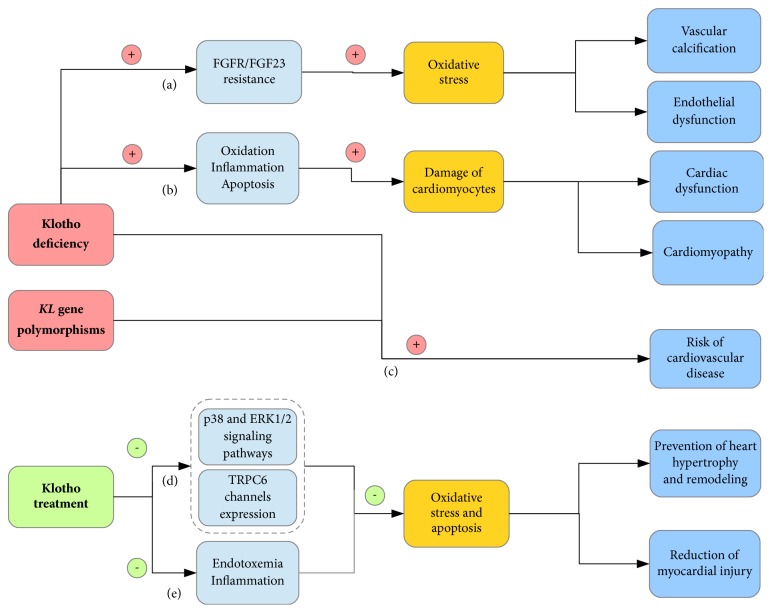
 , induction;
  , reduction.
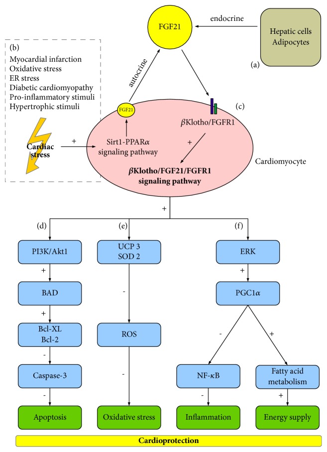
Similar articles
-
[Klotho not only antiageing protein].Przegl Lek. 2017;74(1):25-9. Przegl Lek. 2017. PMID: 29693998 Review. Polish.
-
Klotho Deficiency Causes Heart Aging via Impairing the Nrf2-GR Pathway.Circ Res. 2021 Feb 19;128(4):492-507. doi: 10.1161/CIRCRESAHA.120.317348. Epub 2020 Dec 18. Circ Res. 2021. PMID: 33334122 Free PMC article.
-
Klotho antiaging protein: molecular mechanisms and therapeutic potential in diseases.Mol Biomed. 2025 Mar 22;6(1):19. doi: 10.1186/s43556-025-00253-y. Mol Biomed. 2025. PMID: 40119098 Free PMC article. Review.
-
The impact of preserved Klotho gene expression on antioxidative stress activity in healthy kidney.Am J Physiol Renal Physiol. 2018 Aug 1;315(2):F345-F352. doi: 10.1152/ajprenal.00486.2017. Epub 2018 Apr 25. Am J Physiol Renal Physiol. 2018. PMID: 29693450
-
Role of Klotho in the Development of Essential Hypertension.Hypertension. 2021 Mar 3;77(3):740-750. doi: 10.1161/HYPERTENSIONAHA.120.16635. Epub 2021 Jan 11. Hypertension. 2021. PMID: 33423524 Review.
Cited by
-
Soluble klotho regulates the function of salivary glands by activating KLF4 pathways.Aging (Albany NY). 2019 Oct 2;11(19):8254-8269. doi: 10.18632/aging.102318. Epub 2019 Oct 2. Aging (Albany NY). 2019. PMID: 31581134 Free PMC article.
-
Klotho an Autophagy Stimulator as a Potential Therapeutic Target for Alzheimer's Disease: A Review.Biomedicines. 2022 Mar 18;10(3):705. doi: 10.3390/biomedicines10030705. Biomedicines. 2022. PMID: 35327507 Free PMC article. Review.
-
Association between serum klotho levels and cardiovascular disease risk factors in older adults.BMC Cardiovasc Disord. 2022 Oct 11;22(1):442. doi: 10.1186/s12872-022-02885-2. BMC Cardiovasc Disord. 2022. PMID: 36221064 Free PMC article.
-
Corrigendum to "The Biological Role of Klotho Protein in the Development of Cardiovascular Diseases".Biomed Res Int. 2020 Sep 29;2020:1463925. doi: 10.1155/2020/1463925. eCollection 2020. Biomed Res Int. 2020. PMID: 33062667 Free PMC article.
-
Klotho inhibits IGF1R/PI3K/AKT signalling pathway and protects the heart from oxidative stress during ischemia/reperfusion injury.Sci Rep. 2023 Nov 20;13(1):20312. doi: 10.1038/s41598-023-47686-5. Sci Rep. 2023. PMID: 37985893 Free PMC article.
References
-
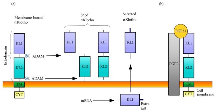
- Matsumura Y., Aizawa H., Shiraki-Iida T., Nagai R., Kuro-O M., Nabeshima Y.-I. Identification of the human klotho gene and its two transcripts encoding membrane and secreted klotho protein. Biochemical and Biophysical Research Communications. 1998;242(3):626–630. doi: 10.1006/bbrc.1997.8019. - DOI - PubMed
Publication types
MeSH terms
Substances
LinkOut - more resources
Full Text Sources
Medical
