

Biomimetic polycaprolactone-chitosan nanofibrous substrate influenced cell cycle and ECM secretion affect cellular uptake of nanoclusters
- PMID: 30671563
- PMCID: PMC6330379
- DOI: 10.1016/j.bioactmat.2018.12.004
Biomimetic polycaprolactone-chitosan nanofibrous substrate influenced cell cycle and ECM secretion affect cellular uptake of nanoclusters
Erratum in
-
Erratum regarding missing Declaration of Competing Interest statements in previously published articles.Bioact Mater. 2020 Dec 4;6(6):1789-1790. doi: 10.1016/j.bioactmat.2020.11.009. eCollection 2021 Jun. Bioact Mater. 2020. PMID: 33336111 Free PMC article.
Abstract
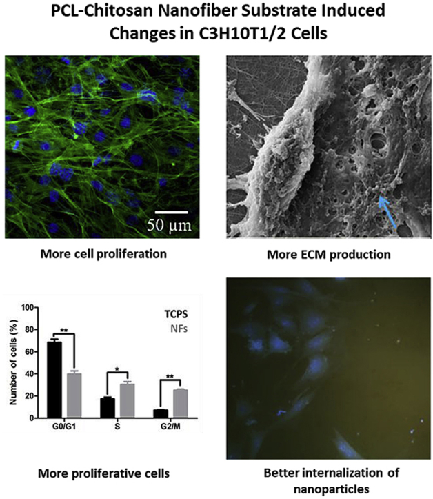
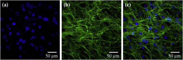
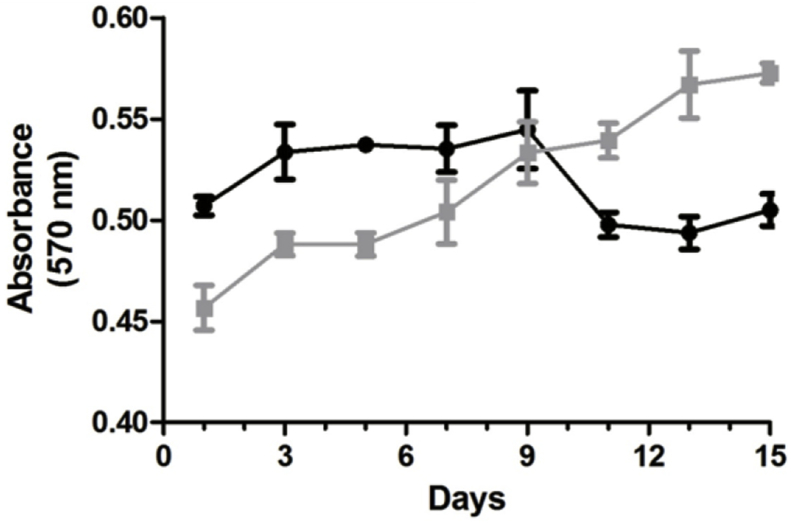
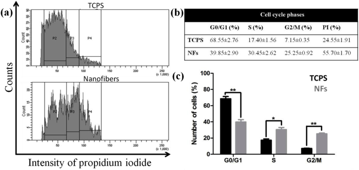
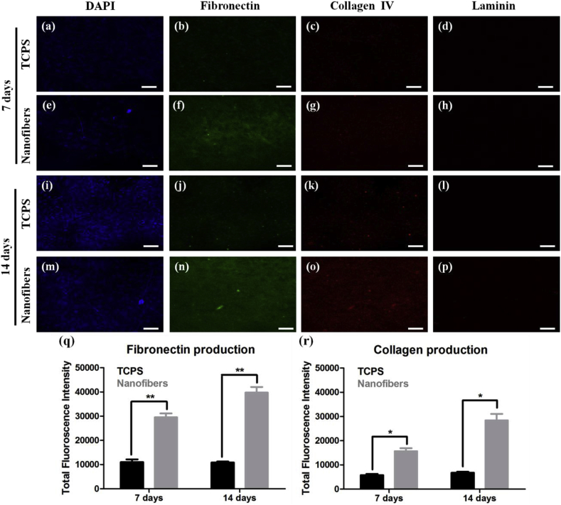
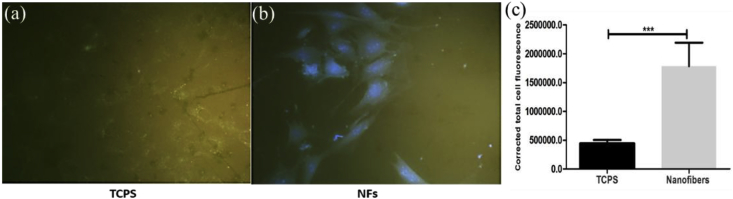
Biomimetic cell culture substrates are developed as an alternative to the conventional substrates. They provide necessary biochemical and biophysical cues to the cells from their surrounding environment for their optimal growth, behaviour and physiology. Changes in physiology of cells growing on biomimetic substrate can essentially affect results of in vitro biological experiments such as drug cytotoxicity, nanoparticle internalization or signalling pathways. As majority of ECM proteins are fibrous in nature, nanofibrous scaffolds have more biomimicking properties. Therefore, in this study, we developed ECM mimicking polycaprolactone-chitosan nanofiber substrate and evaluated its effect on cell morphology, proliferation, cell cycle and ECM production. Further, cellular uptake of BSA-AuNCs has been assessed on conventional and biomimetic substrate in order to demonstrate the effect of these events on cellular properties. It was observed that the cells that were grown for 15 days on the nanofibers, had majority of cells in the proliferative phase of cell cycle compared to TCPS. Moreover, these cells showed extensive collagen and fibronectin production. Due to these conditions C3H10T1/2 cells displayed higher cell internalization of BSA-AuNCs. Overall, this study indicates that the nano-topographical and biochemical environment could alter the cell proliferative behaviour and ECM production, which affects the cell internalization of BSA-AuNCs. Also, PCL-chitosan nanofibrous substrate could be a better alternative to TCPS for cell culture studies.
Keywords: BSA-AuNCs internalization; Biomimetic substrate; Cell cycle; ECM production; Polycaprolactone-chitosan nanofibers.
Figures
References
-
- Pampaloni F., Reynaud E.G., Stelzer E.H.K. The third dimension bridges the gap between cell culture and live tissue. Nat. Rev. Mol. Cell Biol. 2007;8:839–845. - PubMed
-
- Hong S., Kim G. Fabrication of electrospun polycaprolactone biocomposites reinforced with chitosan for the proliferation of mesenchymal stem cells. Carbohydr. Polym. 2011;83:940–946. doi: 10.1016/j.carbpol.2010.09.002. - DOI
LinkOut - more resources
Full Text Sources
Research Materials
