

Abundance of Early Embryonic Primordial Germ Cells Promotes Zebrafish Female Differentiation as Revealed by Lifetime Labeling of Germline
- PMID: 30671659
- PMCID: PMC6441407
- DOI: 10.1007/s10126-019-09874-1
Abundance of Early Embryonic Primordial Germ Cells Promotes Zebrafish Female Differentiation as Revealed by Lifetime Labeling of Germline
Abstract
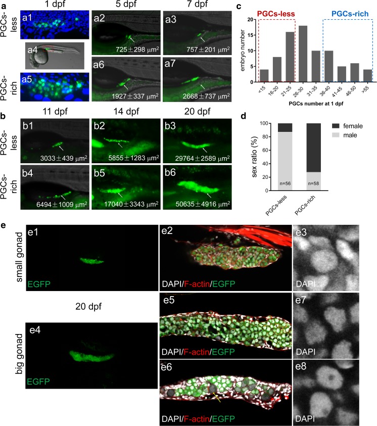
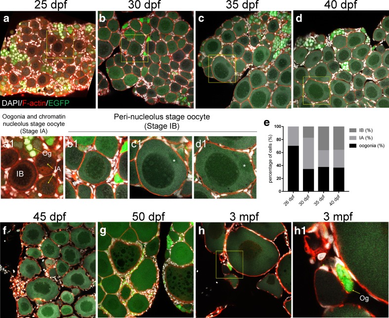
Teleost sex differentiation largely depends on the number of undifferentiated germ cells. Here, we describe the generation and characterization of a novel transgenic zebrafish line, Tg(piwil1:egfp-UTRnanos3)ihb327Tg, which specifically labels the whole lifetime of germ cells, i.e., from primordial germ cells (PGCs) at shield stage to the oogonia and early stage of oocytes in the ovary and to the early stage of spermatogonia, spermatocyte, and spermatid in the testis. By using this transgenic line, we carefully observed the numbers of PGCs from early embryonic stage to juvenile stage and the differentiation process of ovary and testis. The numbers of PGCs became variable at as early as 1 day post-fertilization (dpf). Interestingly, the embryos with a high amount of PGCs mainly developed into females and the ones with a low amount of PGCs mainly developed into males. By using transient overexpression and transgenic induction of PGC-specific bucky ball (buc), we further proved that induction of abundant PGCs at embryonic stage promoted later ovary differentiation and female development. Taken together, we generate an ideal transgenic line Tg(piwil1:egfp-UTRnanos3)ihb327Tg which can visualize zebrafish germline for a lifetime, and we have utilized this line to study germ cell development and gonad differentiation of teleost and to demonstrate that the increase of PGC number at embryonic stage promotes female differentiation.
Keywords: Oogenesis; Oogonia; Primordial germ cell; Spermatogenesis; Spermatogonia; piwil1.
Conflict of interest statement
The authors declare that they have no conflict of interest.
Figures


Similar articles
-
Knockdown of zebrafish Nanog increases primordial germ cells during early embryonic development.Dev Growth Differ. 2016 May;58(4):355-66. doi: 10.1111/dgd.12279. Epub 2016 Apr 29. Dev Growth Differ. 2016. PMID: 27125179
-
Kinesin-1 interacts with Bucky ball to form germ cells and is required to pattern the zebrafish body axis.Development. 2015 Sep 1;142(17):2996-3008. doi: 10.1242/dev.124586. Epub 2015 Aug 7. Development. 2015. PMID: 26253407 Free PMC article.
-
Zebrafish primordial germ cell cultures derived from vasa::RFP transgenic embryos.Stem Cells Dev. 2008 Jun;17(3):585-97. doi: 10.1089/scd.2007.0178. Stem Cells Dev. 2008. PMID: 18576915 Free PMC article.
-
Germ line development in fishes.Int J Dev Biol. 1999;43(7):745-60. Int J Dev Biol. 1999. PMID: 10668983 Review.
-
Avian Primordial Germ Cells.Adv Exp Med Biol. 2017;1001:1-18. doi: 10.1007/978-981-10-3975-1_1. Adv Exp Med Biol. 2017. PMID: 28980226 Review.
Cited by
-
Nanog safeguards early embryogenesis against global activation of maternal β-catenin activity by interfering with TCF factors.PLoS Biol. 2020 Jul 23;18(7):e3000561. doi: 10.1371/journal.pbio.3000561. eCollection 2020 Jul. PLoS Biol. 2020. PMID: 32702011 Free PMC article.
-
Knockout of myoc Provides Evidence for the Role of Myocilin in Zebrafish Sex Determination Associated with Wnt Signalling Downregulation.Biology (Basel). 2021 Jan 30;10(2):98. doi: 10.3390/biology10020098. Biology (Basel). 2021. PMID: 33573230 Free PMC article.
-
foxl2l is a germ cell-intrinsic gatekeeper of oogenesis in zebrafish.Zool Res. 2024 Sep 18;45(5):1116-1130. doi: 10.24272/j.issn.2095-8137.2024.046. Zool Res. 2024. PMID: 39257375 Free PMC article.
-
Bloom syndrome helicase contributes to germ line development and longevity in zebrafish.Cell Death Dis. 2022 Apr 18;13(4):363. doi: 10.1038/s41419-022-04815-8. Cell Death Dis. 2022. PMID: 35436990 Free PMC article.
-
Gonacin: A germ cell-derived hormone with glucogenic, orexigenic, and gonadal activities.iScience. 2023 Sep 28;26(10):108065. doi: 10.1016/j.isci.2023.108065. eCollection 2023 Oct 20. iScience. 2023. PMID: 37860761 Free PMC article.
References
MeSH terms
Substances
Grants and funding
- 31872550/National Natural Science Foundation of China
- 31721005/National Natural Science Foundation of China
- 2018YFD0901205/National Key R&D Program of China
- XDA08010106/Strategic Priority Research Program of Chinese Academy of Sciences
- 2016FBZ03/State Key Laboratory of Freshwater Ecology and Biotechnology
LinkOut - more resources
Full Text Sources
Molecular Biology Databases
Research Materials
Miscellaneous